Ah, a good-natured rant

One of the sources of my daily metaphorical firehose of information that I try to take in is the RSS feed for the blogs at Scienceblogs.

One of the sources of my nearly-daily metaphorical itchy rash of irritation that I encounter along my studies is the fact that as far as most people are concerned (including, most importantly, the people responsible for the required “Microbiology” degree curriculum), “Microbiology” is essentially the study of diseases.

These two collided today when a professor of Epidemiology who is a blogger on Scienceblogs posted offering “Everything you want to know about Microbiology and Epidemiology[…]”. A perfectly reasonable post (downright laudable, actually – it’s part of an effort by several of the bloggers there to be try to get posts going offering explanations of basic concepts in their respective fields). Nonetheless, I couldn’t resist mentioning that what I wanted to know was what it would take to get people to quit assuming microbiology was about diseases (while acknowledging that an epidemiologist was probably not the most sympathetic person to ask…). Hopefully the mini-rant came across in as good-natured a manner as I’d intended.

Those bored enough to care can read it here (the main post is here).

(At least) one of the other organisms…

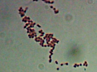
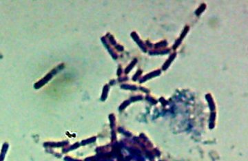
Which I think I shall designate as Ulysses (the Ubiquitous). This sample came from what appeared to be an individual colony on an agar plate made with ~8% salinity diluted Great Salt Lake water. I tried to ‘pick’ a colony with the tip of a hypodermic and ‘poke’ it into the anaerobic tubes. Guess it may have worked, at least with a few cells. (This was done 6 months ago, so if I even got only a few cells over, it’s had plenty of time to grow…) The picture’s a little overexposed (and I tweaked the contrast) though…

I really have no way of knowing for sure, but there does appear to be the same “mass of short rod-shaped bacteria attached to the iron oxide particles” in all of the samples I’ve “live/dead” stained and examined in the microscope so far. So far this is true in every salinity I’ve checked, from about what I estimate to be about 8% salinity (a little over twice as salty as regular ocean water) all the way up to about 27% (pretty much as much salt as you can PUT in water at room temperature and normal pressure). Through the entire range, I see what appears to be the same size-and-shape organism attached to the particles. That would actually be quite interesting. The highest salinity I’ve run into in the “literature” for an organism to be actively growing is about 20%, assuming I did the conversion from “g/L” correctly.
Speaking of which, it turns out the water in the Great Salt Lake is essentially the same as seawater, just saltier (that is, the minor salts in seawater besides “sodium chloride” are also nearly identical in proportion in the lake. There’s just more of it.). Assuming I did THOSE calculations correctly, the amount of sulfate in the water works out to about 0.22M. That’s roughly 8 times as much as in normal seawater – again, assuming my hasty formula-fondling didn’t have a lot of errors.

In any case, there’s a metric [pants]load of sulfate in the water. That means there’s PLENTY of it there for the microbes to breathe and turn into sulfide. No need for any of them to breathe the iron then.

I haven’t given up on that totally though – apparently molybdates can inhibit sulfate respiration, or so I’m told, so I can try making up a solution with some sodium molybdate added. Alternatively, now that I know the Great Salt Lake is very nearly just “concentrated seawater”, I can always make up some “artificial” sea salt, but substitute something else for most of the sulfate and see if things still grow on the iron oxides. That’s likely my next step as a last try to see if any of my Babies can actually breathe the iron. (This is in addition to trying to purify some DNA from Ulysses there, since I think that one is a pure culture.

“Bendy Benjamin” or “Flatulent Fred”?

I was doing a bit of poking around today, and now I’m wondering if the funky-looking spiral/bendy bacteria are of the genus Desulfovibrio. The cell morphology kind of fits – and there’s good evidence that there’s sulfate being reduced down to hydrogen sulfide in the samples (it’s possible there are no iron reducers at all, really, and that the iron reduction is just being caused by reaction with the hydrogen sulfide.) I should mention, though, that this is pretty audacious speculation, since it’s essentially impossible to identify anything based on just looking at it in a microscope…
If nothing else, though, if it IS a “desulfovibrio” type bacteria it might be the most halotolerant one described so far. I wouldn’t be surprised if I’ve actually got a few different sulfate-reducing bacteria growing in there.

That’s something I like about scientific research – even if you don’t get what you’re looking for, you can still find interesting new things (I’ll be a little disappointed if I can find no halophilic iron-reducing bacteria in the samples, but it would still be spiffy to just manage to isolate and identify some previously unknown bug. Or in my currently more-easily-pleased case, just to isolate an odd organism to add to the culture collection I’d like to start…

Hopefully more time at the microscope tomorrow, so maybe I’ll have more (and better) pictures.

“Where Was I?” Femto-episode 20070109 – Astronomy or Microscopy?

I got a chance to check out a bunch of my Gunnison Bay anaerobic iron-reducing cultures in the microscope.

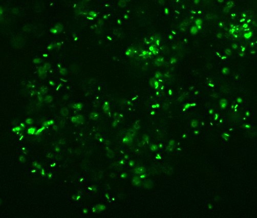
I did a “Live/Dead stain and took a look:


Here you see an image from the Hubble Space telescope of a nebula called…oh, wait, that’s from the microscope. Never mind.

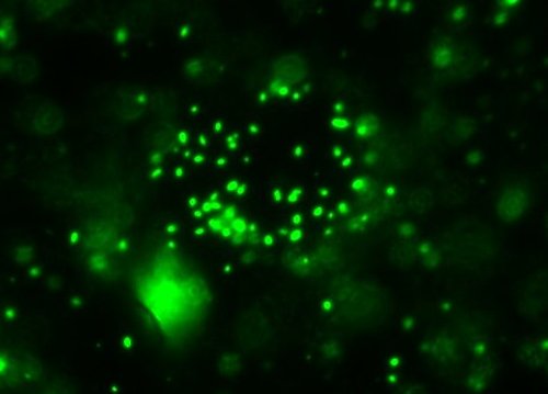
These are the (allegedly) “live” cells. The bright circles are out-of-focus cells – this is a “Wet mount” with huge chunks (from a microscopic point of view) of iron oxides in it, which makes the sample thick enough that it’s hard to focus on more than one part at a time. The vague blur that looks like a gas cloud in a nebula is what you can make out of the chunk of what is more-or-less just wet rust.

This particular specimen is from the positive control of my ongoing experiment (in what I estimate to be roughly 18% salinity, assuming my original Gunnison Bay samples were around 25-26% salinity). If you look close, you can barely make out the bendy/spiral shaped cells almost in focus in a few spots. I believe that this might be the iron-reducing organism that I’ve been trying to isolate. In one of the other samples I checked, I managed to spot one actually actively swimming around, corkscrewing its way around the slide.

I checked one of the experimental specimens that my hypothesis predicts will be growing at least one (and ideally ONLY one) kind of organism and it, too, has a lot of “live” cells growing on the rust-chunks.

I think I shall designate this as-yet-unidentified strain “Bendy Benjamin“…

Other samples from the same place (but not subjected to precisely the same conditions) also seem to have some long straight rod-shaped bugs and some short (straight) rods also still alive in them, so even in the pretty specialized high-salt/no-oxygen iron-oxyhydroxide-being-reduced environment I would guess I’ve got at least three different viable organisms that MIGHT be doing the iron reduction that I’m trying to track down the cause(s) of.

Any questions? No? Does this mean you’re all ready for the quiz?…

“Where Was I”, episode 3 (Now Updated with Lots More Stuff!)

Where Was I, Episode 3 – “It’s a Small World After All” (now updated!)

Study is sucking up most of my time, but that doesn’t mean I never go anywhere any more. Recently, I went to the “Edson Fichter Nature Area”.

Entrance to Edson Fichter Nature Area
Yeah, that place. But look closer.
Marker at the entrance to Edson Fichter
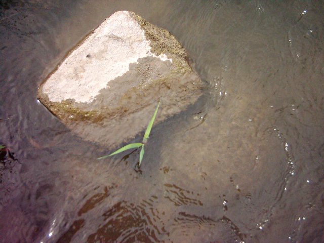
No, no, I mean go inside and look closer. Down there, by the river.
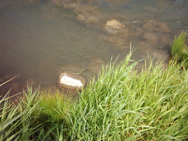
Down by the river
No, no, go closer.

A closer look, down in the river
CLOSER!!!
Microbes!

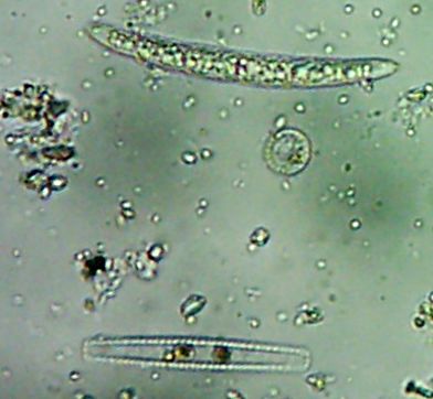
There, that’s a closer look. Incidentally, as you can see, the trick of setting the camera for close-up photos and sticking the little lens opening into the eyepiece of a microscope actually does work. Conceivably, then, the same trick ought to work with any kind of scope, which I’ll have to keep in mind for the next time we go to visit the Bruneau Observatory. But I digress…
long microscopic thing Some really odd thing
I have no idea what all these neat-looking funky things are. Well, not besides the fact that they’re little animalcules with cilia and stuff swimming around and that they’re pretty big as far as microbes go.See, I’m not really into eukaryotes. Needlessly complicated, I say. I’m interested in the cool stuff. Useful stuff.

No ponderous huge protozoans for me, I’m hunting Our Friends, the Bacteria.

See them in there? No, no, you have to look even closer. What do you mean you still can’t see them?

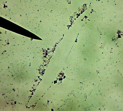
Pretty ciliated thing

Same stuff, now heat-fixed to the slide.  Fire is our friend.All right then, here, let me dry the sample out and heat fix it, then I’ll Gram Stain them for you.Wow. I guess soft, wet, squishy protozoal cell membranes don’t like being heat-fixed, do they? Ouch.

I’m not sure how much of that is bacteria and how much of that is exploded protozoan guts.

Let me look around elsewhere on the slide…
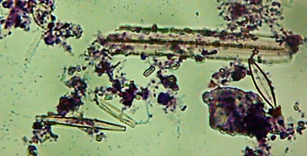
Ghostly, mangled bodies of diatoms litter the landscapeOkay, technically at least some of these things are diatoms rather than “protozoa“.

Kinda neat and creepy at the same time, ain’t it?

Okay, enough staring at hollow shells of dead things, let me see if I can find some bacteria…
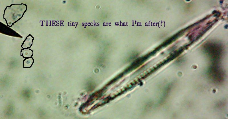
ah, here are bacteriaNow, months later, I’m still not sure how much of the debris on those original slides was bacteria and how much was just crud from the samples. I did, however, get pictures of the 10 isolates that I wanted to try to identify (and of which, as you know from my entry on Willy Bacillus, I so far only know one…)

Of the variety of isolates we got, I pulled out seven that showed some degree of fluorescence under my homebrew UV-LED flashlight, one that was growing in media containing 10mM of a zinc salt, one that was growing in scalding-hot conditions (55°C – which is about the temperature that “melts” collagen into gelatin and makes that tough stew-meat moist and tender), and of course, Willy Bacillus who grew in 10% salt.

Pyotr, Paul, Olga, Ursula, Peggy, Penny, and Ada (the fluorescent bacteria) and Castor (the zinc-tolerant bacterium) all looked more or less like this when I gram-stained them:
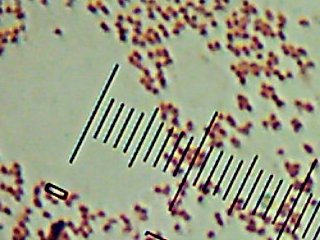
Peggy the Possible PseudomonasPenny the Possible Pseudomonas
I think the Gram-biguous effect is more because of my technique than the bacteria. I believe these are all probably really gram-negative.

There’s a portrait of Willy Bacillus in the other post. Horace was the other interesting-looking culture:
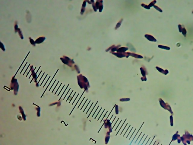
Horace the Hothead
I’m not sure what’s causing the funny spore-like spots or the bulging of the cells. It might be normal, or it might be an effect of the heat-fixing or being in the “cold” (room temperature) or something. I’d be interested to find out what it is.

More fun facts:

  • Olga and Ursula both grew in the ‘fridge (4°C)
  • Ada’s fluorescence was different from the others – it was a dim blue rather than a bright blue-green.
  • Ada also seemed to grow fine on media without nitrogen in it.
  • Peggy was also releasing a green substance into the media.
  • The quick-and-dirty metabolic tests we did (with Enterotubes™ did seem to imply differences between these different isolates.

One lesson driven home this semester was that you just can’t identify bacteria based on simple things like what they look like or a few simple observable traits. However, I still suspect there’s a good chance that everyone but Ada (and maybe her, too) is a Pseudomonas of some flavor, and all of them in any case are probably Gamma-proteobacteria, just based on the fact that when I went looking for information on fluorescent bacteria, nearly everything I found was about species of Pseudomonas, and the ones that weren’t were all still Gamma-proteobacteria. Plus, the media and techniques we used (and possibly the site itself) seemed to heavily favor the isolation of gamma-proteobacteria. If anyone’s bored enough to care, I can post something more about where we got the bacteria and why I wonder if the site had an influence on the heavily slanted taxonomy of our isolates this semester.


And that’s Where I Was.Where Will I Be next? Will my next post be something exciting, interesting, and insightful? Or will I just bore the holy living crap out of everyone with a couple of quickly-crammed-together trips with a couple of minimally interesting pictures? Even I don’t know! Tune in next time and find out!

Meanwhile, as always, questions, comments, and suggestions are welcome.
The famous “Gram Stain”

So far, somewhere between 30% and 50% of this blogs’ current readers have expressed an interest in this, so what the heck. Here, put simply, is why Gram stains work.

Actually, I’ll start with how it works, for anyone unfamiliar with the process, which was invented about 125 years ago. In short:

  1. First, the bacterial sample is put on a slide, allowed to dry, then the slide’s passed over a flame to “heat fix” the bacteria (basically, slightly cooked onto the surface of the slide, like meat stuck to an ungreased hot frying pan)
  2. The sample is soaked with a purple dye (“Crystal Violet”)
  3. The sample is soaked with an iodine solution
  4. The slide is “decolorized” with a quick rinse of alcohol, which should remove the dye from anything that’s “Gram negative”.
  5. Finally the sample is soaked with a pink or red dye to color the “Gram negative” cells so they can be seen as well (unstained bacteria are often nearly colorless so they can be harder to see in a normal microscope.)

“How” is boring, though. What’s interesting is why it works.

Bacteria (with the exception of one group) have cell wall – basically a relatively rigid protein “shell”. The difference between “Gram positive” and “Gram negative” turns out to be that Gram positive bacteria have a thick cell wall, while “Gram negative” bacteria have a thin cell wall, that is also covered by an outer membrane (think of it as a bubble of grease…).

The iodine in the gram stain is a mordant – the combination with the Crystal Violet is “chunky”, and sticks into the cell walls. When the alcohol wash is done, it rinses away exposed Crystal-violet-and-iodine. It also dissolves the outer membrane of gram negative bacteria. Either because there’s not enough cell wall to hold it or because perhaps it sticks in the outer membrane instead, this step washes the dye out of gram negative bacteria, but the gram positive bacteria keep some dye in their cell walls.

The pink/red dye that’s then added actually stains both kinds of bacteria, but you can’t really see it on the gram positive cells because they’re already dyed dark purple.

So, in summary, the Gram stain turns out to be a test for bacterial cell wall types. The results aren’t always reliable (there are “gram variable” bacteria) and it takes some practice to get right (too much alcohol wash and you end up washing the dye out of the gram-positives, too – not enough and some of the gram negatives can look gram positive), but it’s a quick way to start categorizing bacteria.

There, how’s that for some boring science?

Remember “Where Was I, Episode 3?”

Picture of sausage-link-like bacilli

Hi! My name is Willy Bacillus!

“I came from the Portneuf river in Pocatello. I grew in 10% NaCl and I look like slime-coated sausage links. I’m purple in this picture because I’ve been Gram stained. People used to think I was just a strain of Bacillus subtilis, but I’m really Bacillus atrophaeus“.

Yes, I named my bacterial isolates. I figured if we had to give them some arbitrary designation, naming them like hurricanes was just as good as some bizarre system with names like “ZXKUQYB276” or something equally boring.

Unfortunately, we had so much trouble getting the PCR reactions to work we ended up just pooling the class’ sequences rather than having all our own. That means I only got this one. (Actually, it looked like one of my others worked, but not well enough to give enough DNA to sequence.)

I’m trying to talk the instructor into letting me do a 1-credit “Independent Study” session next semester to try to finish getting good sequence identification of Pyotr, Paul, Olga, Ursula, Peggy, Penny, Ada, Horace, and Castor.

The first seven of those all produced fluorescent stuff (probably siderophores, I’m guessing) and while I suspect they’re probably all Pseudomonas species, and almost certainly all gamma-proteobacteria, I was told they actually could be quite different, and I’d really like to find out just how different they really are. Plus, Horace the Thermophile was just kind of neat-looking in the microscope.

Yes…I am a nerd.