“Where Was I?” Femto-episode: “Im in ur kitchen, eatin ur c00kiez”

No, You Can't Have a Cookie - Not Yours

Or: “All Your Bake Are Belong To Us”

Yes, tonight “Where I Was” was in my kitchen.

Introducing “Righteous Fire of Purification Ginger Cookies”, version 1.1! Now with more Culinary Grace! And before anyone asks – no, I don’t have any idea what possessed me to combine internet memes with cooking in this post.

New Features:

  • Reduced pH
  • New Flavor:Lemon Juice!

Source Code: Dry Ingredients

  • 3 Cups of “All-Purpose” bleached flour
  • 1 tsp baking powder
  • 3 Tablespoons (Yes, Tablespoons) Ground Ginger
  • 1.5 Tablespoons (Yes, Tablespoons) Ground Cloves
  • Around 3 teaspoons Cassia Powder
  • A teaspoon or two of Mace (or Nutmeg)

Wet Ingredients:

  • A couple of teaspoons of vanilla extract
  • 2 Large “Grade A” Eggs
  • 0.75 Cups of Molasses
  • A tablespoon or two of lemon juice
  • Not really “Wet” but treated as such:

  • About 1.25 Cups of Unsalted Butter
  • 2 Cups of Granulated White Sugar (Sucrose)
  • About 0.3 Cups of Brown Sugar (Sucrose with a little bit of Molasses added back to it)

How to Compile:

  • Cream the sugars into the butter
  • Add the rest of the “wet” ingredients
  • Add all of the dry ingredients and mix them in thoroughly – add the baking powder LAST (the order of the other ingredients doesn’t matter).
  • Stick the dough somewhere cold to chill
  • Put some baking parchment on a cookie sheet – sprinkle it with sugar.
  • Once chilled, scoop out balls of dough onto the cookie sheet.
  • Sprinkle some more sugar on top of the proto-cookies.
  • Bake 12 minutes at about 350°F
  • Sprinkle more sugar on top of the cookies immediately before they cool.

Installation:

  • Pick up a cookie
  • Insert as much of the cookie into your mouth as will comfortably fit in a manner that would not be considered obscene or freakish
  • Bite off portion of cookie. Chew.
  • Savor the sensations. Be sure to notice and appreciate how the flavors explode on the tongue, like an orgy of purification, preparing your palate for paradise (or at least gratuitous alliteration)
  • Swallow. Repeat the previous two steps until cookie is completely devoured and has become a part of your very being.
  • Visible shudders and audible moans of pleasure and “MMMM!” noises are optional, but recommended.

The only mortal flaw imposed upon these otherwise unmarred cookies is that they keep coming out “crispy-chewy” instead of “soft-chewy” as I want. I was hoping the added acid would help keep the cookies firmer and prevent flattening as they baked – as Tako, the Octopus mentioned in the “Chocolate Chip Cookies” episode – but no luck. Or at least not much. However, as I’d hoped, the lemon juice flavor goes PERFECTLY with the ginger. The texture is a minor flaw (and is actually preferred by some people. Not that there’s anything wrong with that.)

Oh, I know, SOMEBODY is bound to recommend that “shortening” or “margarine” crap instead of butter, since they don’t melt as suddenly as real butter. Feh! Those are An Abomination Unto The Cook! There has to be another way – I refuse to defile these cookies. (Hmmm, how does the melting temperature of lard compare?)

Incidentally, I was completely wrong when I was guessing that the ginger pungency meant it had a mustard-like flavor chemical. It’s nothing of the sort – in fact it turns out that several of my favorite spice flavors have very similar structures – Eugenol (clove), Vanillin (vanilla), Zingerone… All of them have the same sort of “ortho-methoxy phenol” type shape in them. If you look up Zingerone, Vanillin, and Eugenol on Wikipedia, you can compare the chemical structure and see the similarities around the “ring” that I’m talking about.

Oh, and for the internet meme references:

  • See “im in ur base, killin ur d00dz” (Numerous parodies exist – such as This One, though for some reason most of them have cats in them – like this one. (Ran into this one on BoingBoing
  • The picture parodies a picture that appears to be turning into a cliche’ on fark.com. You can see the original here. Incidentally, the photo I used to make this parody comes from “strph” on flickr.com (Original here) released under Creative Commons “Attribution/Share-alike” terms – so if for some reason you can’t resist remixing my masterpiece here…go for it. Just give me credit and allow others to remix yours as well.
  • I’m pretty sure “All Your Base Are Belong To Us” is well covered by the “Internet History” class you have to take to be allowed on the intertubes nowadays, right? No? Well, Wikipedia has the details.

And there you have it – Where I Was™, some food science, and some Internet Nerd History lesson, all in one post. You’re welcome.

“Where Was I”, episode 3 (Now Updated with Lots More Stuff!)

Where Was I, Episode 3 – “It’s a Small World After All” (now updated!)

Study is sucking up most of my time, but that doesn’t mean I never go anywhere any more. Recently, I went to the “Edson Fichter Nature Area”.

Entrance to Edson Fichter Nature Area
Yeah, that place. But look closer.
Marker at the entrance to Edson Fichter
No, no, I mean go inside and look closer. Down there, by the river.

Down by the river
No, no, go closer.

A closer look, down in the river
CLOSER!!!
Microbes!

There, that’s a closer look. Incidentally, as you can see, the trick of setting the camera for close-up photos and sticking the little lens opening into the eyepiece of a microscope actually does work. Conceivably, then, the same trick ought to work with any kind of scope, which I’ll have to keep in mind for the next time we go to visit the Bruneau Observatory. But I digress…
long microscopic thing Some really odd thing
I have no idea what all these neat-looking funky things are. Well, not besides the fact that they’re little animalcules with cilia and stuff swimming around and that they’re pretty big as far as microbes go.See, I’m not really into eukaryotes. Needlessly complicated, I say. I’m interested in the cool stuff. Useful stuff.

No ponderous huge protozoans for me, I’m hunting Our Friends, the Bacteria.

See them in there? No, no, you have to look even closer. What do you mean you still can’t see them?

Pretty ciliated thing

Same stuff, now heat-fixed to the slide.  Fire is our friend.
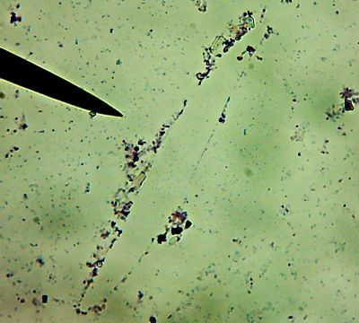
All right then, here, let me dry the sample out and heat fix it, then I’ll Gram Stain them for you.Wow. I guess soft, wet, squishy protozoal cell membranes don’t like being heat-fixed, do they? Ouch.

I’m not sure how much of that is bacteria and how much of that is exploded protozoan guts.

Let me look around elsewhere on the slide…

Ghostly, mangled bodies of diatoms litter the landscape
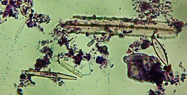
Okay, technically at least some of these things are diatoms rather than “protozoa“.

Kinda neat and creepy at the same time, ain’t it?

Okay, enough staring at hollow shells of dead things, let me see if I can find some bacteria…

ah, here are bacteria
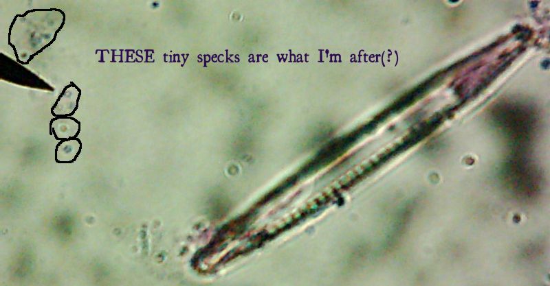
Now, months later, I’m still not sure how much of the debris on those original slides was bacteria and how much was just crud from the samples. I did, however, get pictures of the 10 isolates that I wanted to try to identify (and of which, as you know from my entry on Willy Bacillus, I so far only know one…)

Of the variety of isolates we got, I pulled out seven that showed some degree of fluorescence under my homebrew UV-LED flashlight, one that was growing in media containing 10mM of a zinc salt, one that was growing in scalding-hot conditions (55°C – which is about the temperature that “melts” collagen into gelatin and makes that tough stew-meat moist and tender), and of course, Willy Bacillus who grew in 10% salt.

Pyotr, Paul, Olga, Ursula, Peggy, Penny, and Ada (the fluorescent bacteria) and Castor (the zinc-tolerant bacterium) all looked more or less like this when I gram-stained them:
Peggy the Possible PseudomonasPenny the Possible Pseudomonas
I think the Gram-biguous effect is more because of my technique than the bacteria. I believe these are all probably really gram-negative.

There’s a portrait of Willy Bacillus in the other post. Horace was the other interesting-looking culture:
Horace the Hothead
I’m not sure what’s causing the funny spore-like spots or the bulging of the cells. It might be normal, or it might be an effect of the heat-fixing or being in the “cold” (room temperature) or something. I’d be interested to find out what it is.

More fun facts:

  • Olga and Ursula both grew in the ‘fridge (4°C)
  • Ada’s fluorescence was different from the others – it was a dim blue rather than a bright blue-green.
  • Ada also seemed to grow fine on media without nitrogen in it.
  • Peggy was also releasing a green substance into the media.
  • The quick-and-dirty metabolic tests we did (with Enterotubes™ did seem to imply differences between these different isolates.

One lesson driven home this semester was that you just can’t identify bacteria based on simple things like what they look like or a few simple observable traits. However, I still suspect there’s a good chance that everyone but Ada (and maybe her, too) is a Pseudomonas of some flavor, and all of them in any case are probably Gamma-proteobacteria, just based on the fact that when I went looking for information on fluorescent bacteria, nearly everything I found was about species of Pseudomonas, and the ones that weren’t were all still Gamma-proteobacteria. Plus, the media and techniques we used (and possibly the site itself) seemed to heavily favor the isolation of gamma-proteobacteria. If anyone’s bored enough to care, I can post something more about where we got the bacteria and why I wonder if the site had an influence on the heavily slanted taxonomy of our isolates this semester.


And that’s Where I Was.Where Will I Be next? Will my next post be something exciting, interesting, and insightful? Or will I just bore the holy living crap out of everyone with a couple of quickly-crammed-together trips with a couple of minimally interesting pictures? Even I don’t know! Tune in next time and find out!

Meanwhile, as always, questions, comments, and suggestions are welcome.

The famous “Gram Stain”

So far, somewhere between 30% and 50% of this blogs’ current readers have expressed an interest in this, so what the heck. Here, put simply, is why Gram stains work.

Actually, I’ll start with how it works, for anyone unfamiliar with the process, which was invented about 125 years ago. In short:

  1. First, the bacterial sample is put on a slide, allowed to dry, then the slide’s passed over a flame to “heat fix” the bacteria (basically, slightly cooked onto the surface of the slide, like meat stuck to an ungreased hot frying pan)
  2. The sample is soaked with a purple dye (“Crystal Violet”)
  3. The sample is soaked with an iodine solution
  4. The slide is “decolorized” with a quick rinse of alcohol, which should remove the dye from anything that’s “Gram negative”.
  5. Finally the sample is soaked with a pink or red dye to color the “Gram negative” cells so they can be seen as well (unstained bacteria are often nearly colorless so they can be harder to see in a normal microscope.)

“How” is boring, though. What’s interesting is why it works.

Bacteria (with the exception of one group) have cell wall – basically a relatively rigid protein “shell”. The difference between “Gram positive” and “Gram negative” turns out to be that Gram positive bacteria have a thick cell wall, while “Gram negative” bacteria have a thin cell wall, that is also covered by an outer membrane (think of it as a bubble of grease…).

The iodine in the gram stain is a mordant – the combination with the Crystal Violet is “chunky”, and sticks into the cell walls. When the alcohol wash is done, it rinses away exposed Crystal-violet-and-iodine. It also dissolves the outer membrane of gram negative bacteria. Either because there’s not enough cell wall to hold it or because perhaps it sticks in the outer membrane instead, this step washes the dye out of gram negative bacteria, but the gram positive bacteria keep some dye in their cell walls.

The pink/red dye that’s then added actually stains both kinds of bacteria, but you can’t really see it on the gram positive cells because they’re already dyed dark purple.

So, in summary, the Gram stain turns out to be a test for bacterial cell wall types. The results aren’t always reliable (there are “gram variable” bacteria) and it takes some practice to get right (too much alcohol wash and you end up washing the dye out of the gram-positives, too – not enough and some of the gram negatives can look gram positive), but it’s a quick way to start categorizing bacteria.

There, how’s that for some boring science?