“Live and active cultures” – of beer.

I’ve got a project going to isolate as many yeasts and bacteria as I can from the dregs of a bottle of relatively-famous-brand Lambic ale.

So far, I’ve got at LEAST 3 different types of bacteria and two different yeasts – all of which I suspect are “intentional” – that is, the bacteria are probably lactic-acid bacteria (Lactobacillus, Pediococcus, etc.) which are expected to grow there, and the yeasts I believe to be a Brettanomyces-type yeast and a Saccharomyces yeast (based purely on what I expect to find and the small amount of microscopy that I’ve been able to do so far.)

I have at least one and maybe two different “Gram-positive” rod cultures which I believe to probably be Lactobacillus species. I have several isolates of generic “clusters of Gram-positive coccoids” of which there are at least two different types (which look more or less identical in the microscope, but one of which seems to generate acid while eating mannitol and one that doesn’t).

I have so far named three isolates from Sabouraud agar: Sally, Sid, and Sam.

Sally the Yeast
Sally, the maybe-Brettanomyces-type yeast – 400X magnification (Lactophenol Cotton Blue stain)

Sam the Yeast
Sam, the maybe-Saccharomyces-type yeast – 400X magnification (Lactophenol Cotton Blue stain.)

Sid the [lacto?]bacillus-type-thing
Sid, presumably a Lactobacillus-type bacteria – 1000X magnification (Gram stain)

I’ve also collected four isolates (which may actually just be two different organisms) from an initial inoculation on MSA – BillyBob, JimBob, BettySue, and MarySue. MarySue is the one that seems to be “fermenting” the mannitol.

BillyBob, maybe a Pediococcus?
This is BillyBob (I clipped part of the image and moved it closer to the little “ruler”). The others look essentially the same when Gram-stained.

I’ve also got a bacillus-type (presumably Lactobacillus) critter that showed up on an initial BHI which may or may not be the same as Sid, and I got two more BillyBob/MarySue type colonies on another MRS agar plate.

Interestingly, when I did the original inoculations, it’s the ones that I added the LEAST amount of beer sediment to (20?l) that seems to get the growth – higher amounts may just add so much sugary solution (this stuff is quite sweet) that it inhibits growth.I really hope I can arrange to do molecular analysis (specifically, 16s rDNA sequences) on at least the bacteria, if not the yeast as well. I’d really like to get good identification of these. Assuming they’re real Lambic organisms, they’re probably already in the databases somewhere and should be readily identifiable – assuming someone will let me use up some supplies.

Beer cures flesh-eating bacteria, Staph, Strep, and Anthrax!*

* – These statements have not been evaluated by the Food and Drug Administration. Beer is not intended to diagnose, treat, cure, or prevent any disease, except for maybe hypobeeremia.

No, the title isn’t really true, exactly. However, it does appear to be true that a major component of modern beer – Hops (Humulus lupulus) flowers, really does appear to inhibit “Gram-positive” (Phylum firmicutes) bacteria.


The plates in the picture, clockwise from the upper-left, are inoculated with Bacillus subtilis, Escherichia coli, Pseudomonas aeruginosa (note the green pigment), and Staphylococcus aureus. ON the plates are 5 sterilized paper disks, each soaked with an extract of (again, clockwise from upper-left) Coriander, Hops flowers [Tettnanger], Cassia oil, Clove buds, and Ground Ginger root.

Except for the Oil of Cassia (“Cinnamon oil”), I took 2.5g of each ingredient, boiled it for 15 minutes in distilled water, soaked sterile paper disks in the water, then stuck the disks on top of plates inoculated with the bacteria in question. The cassia oil is about 10?l of the pure, full-strength oil as a sort of “positive control”. At that extreme concentration, it seemed to keep everything away.
The results are even more dramatic than I expected. For one thing, I expected at least some inhibition by the clove extract. The water was the color of a moderately strong tea and smelled strongly of clove, so I would have expected to have enough for some effect…but, no, it was just too feeble. (Had I used pure eugenol, I’d have probably seen the same effect as with the “cinnamon” oil.) Compared to the rest, a mere 15 minutes of boiling a comparatively mild variety of hops flower seems to very effectively prevent growth of certain types of bacteria – which would presumably include the varieties mentioned in the title of this post.

Hops skin-lotion to appear at hugely inflated prices on health-food-store shelves in 3…2…1…

Incidentally, if it does, I wouldn’t use it. “Gram positive” bacteria make up a substantial portion of the “normal flora” of healthy skin. Killing them off might easily leave room for other bacteria to take over and cause problems.

It does make me wonder about other possible uses of this effect, but I’ll save that for another time.

I’ll close by pointing out how useless allegedly “anti-bacterial” spices seem to be by comparison. Kind of puts the whole ridiculous notion of medieval cooks using spices to inhibit spoilage or to treat “rotten” food in its place, I’d say. It also implies that hops isn’t going to prevent “spoilage” of beer by itself, given that (for example) vinegar bacteria aren’t “gram-positive” types, nor are all the lovely ?-proteobacterial butt-bacter organisms like E.coli going to be affected…at least not by the hops. More experimentation to be performed at some later date.

This is just a simple experiment on the side of the main one I’m performing, where I attempt to isolate as many different viable organisms from a bottle of famous-brand Belgian Lambic ale as I can, hopefully for use in other foods (sourdough? Yogurt? And, of course, beer…) later.

Chunky Bacon Agar, and Expired Jell-O™ again

I’m still working on the “Taxonomy of Yogurt” post which I currently plan to do next, but I’m overdue for a post already – therefore, here’s a brief one to keep my legion of adoring fans appeased until the next long post, here’s a short one.

Part 1: I got an interesting search-query hit recently – looks like (I’m guessing) a technician working at a famous pharmaceutical/healthcare-product company ran into the same problem I did during my current Bacterial Virology lab – “chunky microbiology agar in microwave”.

Agar is nifty stuff to use for microbiology. Dried, it’s a lumpy powder. To use it, you dump around 1-2% w/v (more or less, depending on the consistency of agar that you need) into water and heat it up to dissolve it. It’s basically seaweed-JellO™ – except it’s not actually affiliated with Kraft Foods nor made of gelatin. Anyway – once it’s dissolved, it’ll cool into a gel.

The nice thing is, you can make up a bottle of this stuff and let it solidify, and store it (sealed) for quite a while. When you want to use it, you can just stick it in a microwave oven to melt it back down. It has to get pretty hot for this, but it then stays liquid until it gets down nearer to room temperature, so you’ve got plenty of time to pour it into plates or tubes or whatever.

For bacterial virology purposes, we make up a “soft agar” (about 0.8% agar, as I recall) to make an “overlay” – after mixing bacteria and virus together into a small amount of melted [but mostly cooled, so it doesn’t fry the bacteria] agar, we pour the soft agar in a thin layer over the top of a regular layer of nutrient agar in a plate. (The idea is that then wherever there is a virus that can infect and kill bacteria, it’ll wipe out all the bacteria growing in a particular part of the overlay, leaving a cleared “plaque” – you can then count how many plaques there are to find out how many virus were in the original sample, for example).

Earlier this semester, we had a fair amount of trouble with this. We’d go to pour the overlay and it’d come out chunky, even though it looked completely melted when we prepared it. Fortunately, the problem is simple and easily solved – you just need to nuke the heck out of the stuff, frequently swirling the container to make sure it’s completely mixed. What seems to be happening is that a few bits of agar remain unmelted but hard to see if you’re not careful, and those bits allow the melted agar to coagulate around them more readily. In short, the trick is to make really sure that all of the agar is completely melted.

Note that you have to be careful while doing this – lots of bubbles end up coming out of the agar when you swirl it, and it can easily foam out of the container and burn your hand. (Oh, obviously you also need to leave the lid a little loose to let off the pressure.) Of course, the stuff will be really hot when you’re done with the microwave, but as mentioned before, it’ll stay liquid until it is much cooler before it solidifies. If you set the bottle in a warm-water bath (~50°C or so) you can basically walk away for hours, leave it overnight, or whatever, and it should still be completely liquid and smoothly pourable – not to mention cool enough to handle with bare hands – when you get back.

And on the subject of gelled material – the fact that I mentioned all the hits about expired Jell-O™ in the previous post seems to have substantially increased the number of “expired-JellO™-related” hits I’ve gotten, so here’s a slightly more expanded update.

Assuming one is referring to the “instant gelatin” powders (regardless of brand), as far as I can tell they ought to be safe to use almost indefinitely. Officially, Kraft Foods, the owners of the Jell-O™ trademark say that the expected shelf-life is 2 years (“24 months”). I still think, personally (Note – Your Mileage May Vary, Do Not Try This At Home, and other standard disclaimers apply here) just like sugar, that it is probably safe to use practically forever as long as it doesn’t get wet (and isn’t stored in humid conditions). I don’t think anything of consequence would be able to grow on the dry powder, and I find it unlikely that the normal flavorings would be prone to suddenly become poisonous as a result of ordinary aging. The only thing you might have to worry about is maybe some of the flavoring compounds getting slowly oxidized by the air, so maybe the result wouldn’t taste quite the same. As far as I am concerned, so long as there weren’t fuzzy clumps growing in it, if the contents of the packet were still flaky/powdery, I’d most likely go ahead and use it, and not expect to suffer any ill effects.

‘course, if you read my obituary someday and it notes that I died of expired-gelatin-poisoning, you’ll know I was wrong…

UPDATE: I empirically test the toxicity of of expired JellO® on my own body! The saga begins here!

I’ve got DNA! I’ve got DNA! I’ve got DNA!…

As you can see, I’ve got DNA. I’ve been trying to get this stuff successfully extracted and the 16s rDNA amplified for months (off and on) now. Looks like doing the whole-genome-amplification step first did the trick – this is from a set of mixed halophiles in a phlogisticated environment growing in approximately 18% salt solution, and they grow very slowly. It’s hard to get enough DNA extracted from such a small population to do useful work with.image of electrophoresis of 16s DNA amplicons

The gel “bands” you see to the right of the image are (or at least should be) made of copies of the DNA which codes for various “16s small-subunit ribosomal RNA” sequences for the one-or-more different kinds of prokaryotes living in my culture. The brighter the band, the more DNA is there.

Since all of the samples were processed exactly the same way, then, the brightness of the band should, at least indirectly, indicate how many bacteria were in each sample to begin with. This isn’t necessarily true – there can be variation in how many copies of the gene each kind of bacteria has, so if the populations are very different the results could be misleading. Still, it’s gratifying that my little ‘proof-of-concept’ experiment not only finally gave me some DNA but even shows exactly the kind of difference I originally hoped for. (The second “lane” from the top with the brightest band was SUPPOSED to be enriched for certain types of bacteria, according to my hypothesis. The first “lane” should have had less, and it does. The third lane is my “positive control”, growing without special influences, and the fourth lane with no DNA visible is my negative control, which I hoped would have little or no DNA (indicating little or no bacteria growing in it) – and that’s what I see.

It doesn’t prove anything at this point, but finally getting results and having them turn out to look the way I’d hoped is a good start. I wonder if I can get them into a clone library, separated, and sequenced before next weekend?

I’ll have to remember to thank last semester’s “Senior Seminar in Microbiology” instructor for assigning me that paper[1] – I thought some of the technology described in it sounded like it’d be useful to me personally.

Anybody else going to the Northwest Regional ASM meeting next weekend?…

[1] Wu L, Liu X, Schadt CW, Zhou J: “Microarray-based analysis of subnanogram quantities of microbial community DNAs by using whole-community genome amplification.” Appl Environ Microbiol. 2006 Jul;72(7):4931-41.

Officially “Gram-positive” – the Firmicutes

In a typical student microbiology lab, it seems whenever you get your hands on some bacteria the first thing you do is check to see if it’s “Gram-positive” or “Gram-negative”. But does this old Victorian-era test still mean anything useful in the context of modern bacterial taxonomy?

It would seem that it actually does. In my admittedly limited experience, an un-ambiguous Gram-positive result using the standard procedure nearly always indicates bacteria in the phylum firmicutes, sometimes also referred to as the “low G+C gram positives”.

The “low G+C” part has to do with the chemical characteristics of the DNA. If you’re already familiar with DNA’s structure then you probably already know what this means, but for everyone else, in brief:
DNA is made of strings of four different chemical “bases” chained together. The bases are “Adenine”, “Guanine”, “Cytosine”, and “Thymine” (abbreviated as A, G, C, and T). These bases make up a sort of chemical “alphabet”, which encode how to make various proteins. These chemical bases each have an opposite base that they are attracted to – Adenine to Thymine, Cytosine to Guanine, so DNA’s strings end up matched with an “opposite” string, which together form the easily recognized “double helix” shape of the overall DNA molecule. The relevance here is that the attraction between the Guanine and Cytosine (“G+C”) is stronger than the attraction between Adenine and Thymine, so you can estimate relatively how much Guanine and Cytosine is in an organism’s DNA by how tightly the two strands of DNA stick together.

And, yes, there is a “high G+C” gram-positive group, with a larger proportion of the G and C bases as compared to the A and T. That’s the “Actinobacteria”, which includes the “acid-fast” Mycobacterium group. But that’s a topic for another post.

The firmicutes, with the exception of one group that I know of, all have a distinctive, simple outer structure. It’s something like this:

Imagine a water balloon filled with lime Jell-O®. Now, tightly wrap the water balloon with a piece of thick, padded packing blanket. The thick layer of packing blanket is the cell wall. The balloon is the inner cell membrane. The Jell-O® is the cytoplasm. (It’s Lime merely because I like lime Jell-O®.) It might be worth noting that since this group of bacteria relies so much on it’s cell wall, they are more likely to be killed off by the ?-lactam antibiotics (which specifically attack bacterial cell wall generation) than other types of bacteria.

There is one exception to this structure – the class of firmicutes known as the mollicutes. There’s one example of this group that gets mentioned in basic medical-centric microbiology classes: the genus Mycoplasma (as in Mycoplasma pneumoniae). Members of this class have lost their ability to make cell walls entirely.

By contrast, a typical “Gram-negative” cell has a more complicated outer makeup. In brief, start with the same lime Jell-O® balloon, but instead of a thick packing blanket, wrap it with a single thin layer of cloth, and then stuff the whole thing inside of another lime Jell-O® balloon. You can also imagine a bunch of valves stuck through the outer balloon to let stuff in and out, but then you end up imagining the Jell-O® spurting out all over the place and the whole analogy breaks down. If it helps, you can also imagine that if you punched out a section of a gram-negative outer structure with a cookie-cutter, you’d get something kind of like an inverted Oreo® cookie, with easily-dissolved filling on either side of a thin layer of cookie. Anyway, the outer balloon represents the outer cell membrane, the cloth is the (much smaller) cell wall, and the layer of lime Jell-O® between the outer balloon and the cell wall is called the “periplasmic space”.

Interestingly, despite this difference in complexity, the molecular evidence[1,2] seems to indicate that the simpler firmicutes diverged evolutionarily from the more complex “gram-negative” types, not the other way around. The lineage suggested by the 16s gene sequences implies that both types of Gram-positives split off from the Gram-negative type bacteria as a single ancestral group, and some time later the two types diverged into separate groups. If I had to bet, I’d personally put my money on the evolutionary sequence going Gram-negative-type->actinobacteria->firmicutes.

Another characteristic of some (but not all) of the firmicutes is the production of “endospores”. Unlike the spores of molds or Myxobacteria, endospores aren’t reproductive. Instead, they’re a like a lifeboat, or perhaps a metaphorical bomb-shelter in the cells’ also-metaphorical basement. If environmental conditions get unpleasant, the bacteria essentially pull their DNA and a few necessary enzymes into a small, thick, multilayered compartment – the endospore – where they can wait, protected and dormant, until conditions become comfortable again.

The special “Endospore stain[3]” uses a dye called Malachite Green and works somewhat similarly to the Gram and “Acid-fast” stains – the extra-thick spore coats retain the green stain (once it’s been driven in by some extra heat) while the decolorization rinse washes it out of everything else. I still need to try using a microwave oven for the heating step…but that, too, is a topic for another post.

If your microbiology class was typical, when you think of the firmicutes, you may think of little more than “Strep throat”, “Anthrax”, and “MRSA“. If so, though, you’re missing out on the useful ones.

Since Gluttony is my second most favorite “Deadly Sin™”, I tend to think of food-related possibilities here. And, no, I don’t mean Botulism.

Yogurt and Kumiss and Kefir, Sauerkraut and Kimchi, Sourdough bread, Salami, and Belgian Lambic ales all involve (at least in part) growth of various lactic acid producing firmicutes. Mostly members of the Lactobacillus genus, though if you take a look at the labels of some of the “Live and Active Cultures” yogurt you may also spot a close relative of the ‘Strep throat’ bacterium in the list. These kinds of bacteria can be happy members of the “Normal Flora” of the healthy human gut.

Another, more obscure example is “Natto”. A strain of Bacillus subtilis, originally derived from rice straw, is allowed to ferment soybeans. The end result is a pungent mass of beans, covered with gooey slime, and having an odor vaguely resembling old cheese. I’ve actually eaten it, and they aren’t nearly as bad as this makes them sound…though I personally still haven’t acquired a taste for them. You may have seen this stuff if you watch Iron Chef – it was used as the Secret Ingredient in one episode. There does appear to be some real potential for it as a “health food”, though, which you can see if you poke around Google or PubMed searching for “Natto”. Maybe next time you end up in the basic Microbiology lab and you’re given some B.subtilis to look at you can whip out a jar of soybeans to inoculate while you’re at it. (Note that I wouldn’t actually recommend eating the results in this case…)

Comments and corrections, as always, are welcome.

P.S. no affiliation with any of the trademarks mentioned should be inferred here – I just figured the trademarked names would be more recognized than terms like “gelatin” or “sandwich-cookie”…

[1]Sheridan PP, Freeman KH, Brenchley JE: “Estimated minimal divergence times of the major bacterial and archaeal phyla.” Geomicrobiol J 2003, 20:1-14.
[2]Battistuzzi1 FU, Feijao A, Hedges SB: “A genomic timescale of prokaryote evolution: insights into the origin of methanogenesis, phototrophy, and the colonization of land.” BMC Evolutionary Biology 2004, 4:44
[3]Schaeffer AB, Fulton MD: “A simplified method for staining endospores.” Science 1933 77:194
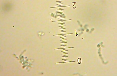
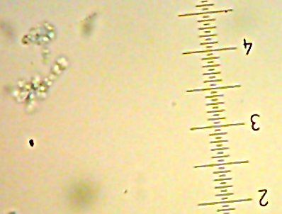
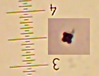
How does one categorize prokaryotes?

If you’re taking or have taken only basic or “medical” microbiology courses, it may seem like bacteria are still classified by ancient 19th-century criteria. Are the bacteria Gram-positive, Gram-negative, Acid-fast? And then, are they round, straight, or bendy? Then, to categorize any further, there’s a bewildering array of culture tests you can go through – some of which seem archaically specific with quaint un-intuitive terminology for the results.

Take “hemolysis“, for example. Considering how wide-ranging the environments are in which different bacteria can live, going through all that effort to see if the bacteria secrete any special substances that have a special effect on sheep red blood cells seems less than helpful. Okay, in fairness, when you’re dealing specifically with disease bacteria with the potential to possibly get into somebody’s bloodstream this test is useful, but in the entire population of all bacteria, this is a ridiculously tiny number of species.
And then there’s the terminology for this one – if the blood cells in the media are popped open but not totally destroyed (the media underneath will be greenish from the iron compounds released from the popped cells) it’s called “?-hemolytic”. If they ARE essentially totally destroyed (the media underneath the bacteria clears completely) it’s “?-hemolytic”. Not bad so far, but what if the bacteria doesn’t visibly destroy the blood cells at all? Why, that’s “?-hemolytic”. No, not “non-hemolytic”, even though that’s what it means. And, no, I have no idea why this is. I’m glad whoever came up with this scheme wasn’t a legislator. Otherwise, people who were accused of being murderers but were proven to be innocent would be declared to have committed “?-homicide”…

Using these kinds of phenotypic tests does have some advantages – most of them have been around so long that they’ve been thoroughly tested, they’re usually pretty easy to do, and if you’re hiring people to do bacterial classification grunt-work, finding people who can handle “mix this stuff together and see if it gets clumpy” is easier and cheaper than finding people to do more complex molecular work. On the other hand, there are some major disadvantages.

Firstly, there doesn’t seem to be any publically-accessible data repository with useful information of this kind that I’ve been able to find, unlike, say, the genetic data readily accessible at Genbank or the RDP-II. Sure, you can sometimes find some of this kind of data in papers describing specific species or strains, but to use this kind of information you have to already have a good idea of what your microbe might be – you can’t just plug “it ‘ferments’ lactose, sucrose, and xylitol, but not sorbitol or fructose, it’s catalase-negative, liquifies gelatin, and grows happily even when there’s tetracycline in it’s culture media” into a decent online database anywhere that I’ve been able to find and get a listing of known possibilities.

The other problem is that a lot of these kinds of traits aren’t necessarily part of the bacterium’s own genome. Bacteria often inherit certain traits from “bonus” DNA that they can pick up from other bacteria (“plasmids”) or from a viral infection. Yes, bacteria can be infected by viruses. Resistance to antibiotics is commonly spread by plasmids (though not always – I seem to recall that Klebsiella pneumoniae, for example, has a penicillin-resistance ability that actually is part of its core genome.) A number of diseases are actually caused by bacteria getting infected with viruses. Maybe your case of Necrotizing Fasciitis isn’t because your Streptococcus pyogenes on your skin, but because your Strep. pyogenes is rabid

The most reliable way available to work out who a microbe is involves comparing versions of a gene that essentially is never moved around between different microbes, but all microbes have. For prokaryotes, the gene in question is the “16s small subunit ribosomal RNA” gene sequence – or just called “16s“. This is what the taxonomy you can find at NCBI for bacteria and archaea is based on.

You know, when I started this post, I was just going to say that I wanted to get a more intuitive understanding of prokaryotic taxonomic groups and to that end was going to put together some posts on particular types of bacteria and archaea. I hope all the “bonus” background that seems to have come pouring forth from my overheated mind is useful.

Anyway, stay tuned this weekend for a post on firmicutes at some point.

Making the great leap out of the 19th Century…”Acid-Fast” staining

The “standard” acid-fast differential staining process for the “High G+C Gram-Positive” bacteria[1] as we learned it is pretty archaic.

It goes something like this:

  • Smear and heat-fix the slide
  • flood the slide with Carbolfuschin
  • Heat the slide for 5 minutes in steam over a boiling water bath
  • Rinse with “Acid Alcohol
  • Stain for a minute with Methylene blue.

At the end, anything “Acid-Fast” (having a “waxy” outer layer) will show up red, anything else will be blue.

My objection is the messy and time-consuming steam-bath.

Today’s lab included one “unknown” which we suspected to be a Mycobacterium, so while one of us was going through the tedious 19th-century-style procedure, I decided to try something.

The steambath heat is just intended to (I believe) slightly “melt” the waxy layer of the cell and otherwise help “drive” the dye into it. So, instead of dealing with the time to make a water bath, heat until it steams, and then wait for the slide to sit there and hope the bubbling bath doesn’t splatter the slide with crud, I just stuck the flooded slide in the lab microwave and cooked it for 20 seconds.

It worked. Quite well, actually (other than letting the slide dry out, leaving some crystals on the slide) – the bright red mycobacterial cells showed up nicely. I’m annoyed that my ‘stick the camera up to the eyepiece’ technique came out slightly out of focus (I may see if I can enhance it later – if so, I’ll post it.). Somebody commented that it looked as good as a “textbook” example, which was nice for my ego…

Unfortunately, I guess I’m far from the first person to think of this. I don’t know if anyone’s done this exactly the same way, but This procedure describes directly heating the Carbolfuschin in the microwave and soaking the slides directly in it. There is also apparently an old Lancet[2] article which I don’t currently have access to – I’ll have to check it out later.

Next thing to do is try the endospore stain this way. Behold, the miracles of applying last century’s technologies to the problems of century-before-last!

[1] Ehrlich P. Zur Fa¨rbung der Tuberkelbakterien. Aus dem Verein fu¨r innere Medizin zu Berlin. Deutsche Med Wochenschr 1882; 8:269?270
[2] Hafiz, S., R. C. Spencer, M. Lee, H. Gooch, and B. I. Duerden. 1984 . Rapid Ziehl-Neelsen staining by use of microwave oven. Lancet ii:1046.

Poisoning Prokaryotes in the Park

(Well, okay, it was a “Pathogenic Microbiology Lab”, not a “Park”, but whatever).

Antibiotic Susceptibility of a Poor, Innocent Microbe

Objective:Experience the awesome power of the mighty Antibiotic Susceptibility Test, wielded against an unsuspecting bacterial organism!

Introduction:

Every day, billions of innocent bacteria are ruthlessly slaughtered by antibiotic substances introduced into their callous, inconsiderate hosts. Condemned to death as nuisances due to nothing more than the potential inconvenience of debilitation, tissue necrosis, death, halitosis, and other minor problems, the unstoppable might of all medical science is focussed on our prokaryotic friends like medical professionals around the world focussing the rays of the sun through a thousand magnifying glasses to obliterate innocent prokaryotic “ants”.

The ?-lactam antibiotics – the blahblahcillins (Penicillin, Ampicillin, etc.) and the Cefablahblah compounds (Cephalosporin, Cephalothin, Cefuroxime, etc.) all interfere with the formation of the bacterial cell wall – loosely analogous to the human epidermis. This treatment viciously targets the hardest-working, actively-reproducing bacteria, spilling their guts as they attempt binary fission, while leaving the lazy, dormant microbes alone. Gram-negative bacteria are somewhat protected from this torture by their outer membrane, but some (such as ampicillin) can affect even some of them. Gram-positives, with their simple structure, are hardest hit. A few microbes have learned to counter this by secreting an enzyme which disables many of these drugs, though medical science has countered with clavulanic acid – an inhibitor of the ?-lactamase enzymes. Enzymes resistant to inhibition by clavulanic acid are being developed as part of this continuing arms race.

Chloramphenicol is an artificially manufactured bacteria-poisoning chemical synthesized in laboratories (though it was originally obtained during interrogation of a captured Streptomyces species) which interferes with protein synthesis at the 50s ribosome. Erythromycin has the same affect, by a slightly different mechanism, and is a macrolide – a class of large molecules with lactone rings which resemble in shape the poisoned ninja throwing-stars seen in movies. Both of these chemicals are bacteriostatic rather than bacteriocidal, but are broad-spectrum. Chloramphenicol’s devious action sometimes backfires on a small number of people, causing potentially fatal aplastic anemia.

The Sulfonamides are also bacteriostatic, and are competetive inhibitors of enzymes that convert the nutrient PABA into biochemical products vital for prokaryotic health. Much like a hypoglycemic person with diarrhea given a “cupcake” made entirely out of Olestra® and Sucralose, the microbial victim of this chemical ingests it but finds that it merely inconveniences the metabolic processes rather than feeding them.

Tetracycline (the first of the Tetracycline-type antibiotics) and Tobramycin (an Aminoglycoside) both jam the gears of protein synthesis, inhibiting the action of the ribosome in the former case, and actively causing erroneous protein formation in the latter. The latter effect is outright bacteriocidal, causing the poor bacterium’s protein assembly systems to make broken enzymes until the cell’s protein factory is bankrupt and has to lay all the enzymes off. Tetracyclines appear to only slow down the cell, but in the cutthroat competition for cellular activity in the human body, this stumbling can be a death sentence for the business of prokaryotic replication.

In order to determine which of these lethal agents to deploy against the oppressed bacteria, a medical professional may capture a microbe and torturously test various agents on it, watching without emotion to see which ones destroy the microbe most efficiently. This awful, coldly clinical process is standardized in the Medical Microbiologist Field Manuals on Interrogation as the “Kirby-Bauer”[1] antibiotic susceptibility test. In these tests, 6mm diameter paper disks soaked with various antibiotics in specific amounts, is pressed onto a growing young microbe culture to see which ones are most destructive, leaving desolate areas devoid of life in the culture…

Recently, we got to do this..

Materials and Methods:

A colony of Pseudomonas aeruginosa was lured into a culture tube with the promise of free candy. Happily replicating, this culture was transported to a secret location containing Mueller-Hinton agar. 100?l of this culture was told that it had won an all-expense-paid stay at a four-star hotel with room-service, and was plated onto the agar. The culture was then strapped down and tortured for its secrets by application of 12 antibiotic-soaked disks. The torture was performed at 37°C for 24 hours, and the results measured with a ruler.

Results:

The defiant Pseudomonas organism bravely withstood the application of Ampicillin, Cephalothin, Chloramphenicol, “Triple Sulfa”, Nafcillin, Cefazolin, Amoxicillin with clavulanic acid, Cefuroxime, and Penicillin G. Erythromycin seemed to cause the subject some discomfort. It was not quite able to grow to the edge of the antibiotic disk all the way around but was able to get within less than a millimeter of it, even touching it in a few spots. Tetracycline was unbearable to the subject, who was limited to a 11mm (diameter) zone of inhibition around the Tetracycline-containing disk. Finally, Tobramycin (zone of inhibition approximately 23mm) finished breaking of the subject, who then confessed to several murders of immunocompromised individuals, robbing a “Wal-Mart”, and once molesting an archeaean of the genus Thermoplasma. Investigations may already be underway to determine the authenticity of these confessions. Then again, they may not.

Conclusions and Discussion:

Pseudomonas aeruginosa is a hardy little bugger who can put up with a wide variety of antibiotic insults. Its weak point appears to be its ribosomal machinery, as this was the target of the three drugs which had any apparent effect. Should intelligence indicate the threat of attack by the terrorist Pseudomonas organization, ribosome-targeting, protein-synthesis-inhibiting agents should be deployed as a countermeasure.

References:

[1] – Bauer AW, Kirby WM, Sherris JC, Turck M, “Antibiotic susceptibility testing by a standardized single disk method” Am J Clin Pathol. 1966 Apr;45(4):493-6

[2] –Tortora GJ, Funke BR, and Case CL, “Microbiology – An Introduction (sixth edition)” 1997, Benjamin/Cummings Publishing Company, Menlo Park, CA

Cheap Miscellany

Tonight’s post will be a bit short, but I’ll try to make up for it tomorrow. (Three more days of Just Science week to go…)

First, a quick unsolicited plug for someone else’s site: Aetiology is doing a series of posts on “Normal Flora” – that is, the microbes that normally live on and in healthy people (as opposed to microbes that just cause disease). Since that’s an area where my own interests overlap with the more conventional “medical” microbiology, it seems appropriate to mention it here. As of right now, there is a Part 1 and a Part 2. Interesting and informative stuff.

Second, a small addendum to the previous post – I mention that I think when people say “Gram-Positive” they generally really mean Firmicutes, but I just realized there’s one exception to that. The Firmicutes actually include Mycoplasma and related bacteria – which have no cell wall at all. I would guess their ancestors were normal “Gram-Positive”-type firmicutes but somewhere along the way “lost” function of a key gene involved in making the thick cell wall. (There’s a similar group of Archaea – the Thermoplasmata. ) These don’t stain Gram-positive (and perhaps don’t stain gram-negative, either – seems like such fragile things would be destroyed by the staining procedure). In order to see these in the microscope, the standard method seems to be to use a chemical that actually stains DNA instead. Instead of staining the outer surface of the cell like most of the classical stains, this stains the inside of the bacteria (which tend to have their DNA spread more or less throughout the entire cell in one form or another, since they have no nucleus to pack it into) with a fluorescent material, which you can then see in the microscope with the right kind of light. You can also use this kind of technique for other bacteria, too. The “Live/Dead” stain I previously mentioned works this way, if I remember correctly.

Since you probably don’t want to try heat-fixing Mycoplasma, you have to use a “fixative” (a preservative made of alcohol and pure acetic acid) and then air-dry the slides.

This leads to one last correction – in a previous post I suggested that it was unlikely that you could “glue” your smear to the slide rather than heat-fix (assuming that the “glue” would interfere with the subsequent staining and viewing of the sample) – this is actually not completely correct. I heard today of a protocol for doing endospore and acid-fast stains which called for mixing the culture sample with serum on the slide to make it stick, so there are at least a few ways to “stick” cells to the slide and still look at them.

Are “Acid-Fast” bacteria Gram-positive or Gram-negative?

I was wondering what today’s post ought to be – but a Google™ search that reached the page posed an interesting question and made it easy.

Someone from a Miami, Florida college got to this site after asking Google:
“Assuming you could stain any cell, would an acid-fast [bacterium] be gram-positive or gram-negative?”

Therefore, today’s post will deal with some Microbial Physiology.

The easy answer is, of course, “no“.

A more useful answer, though, is that it depends on what you mean by “Gram-positive”.

If one takes the “Assuming you could stain any cell” part of the question to mean that you’ve done whatever it takes to get the Crystal Violet/Iodine into the cell wall, and you mean “will the cell still look purple instead of pink at the end of the Gram Stain process”, then I’m pretty sure the answer would be yes, that it would be “Gram Positive”. It actually IS possible to stain “acid-fast” bacteria with the Gram stain. The catch is that it takes 12-24 hours of staining (according to Gram’s original paper) rather than a minute or so. This still counts as Gram-positive, though, and in fact the whole Phylum of Actinobacteria (including the Mycobacterium genus) is considered “High G+C Gram-positive”. (If the query was for an exam or homework problem, this is probably the answer you’re looking for.)

On the other hand, if you mean “does the cell wall structure of an acid-fast bacterium better resemble Gram-positive or Gram-negative bacteria?”, you can make an argument that instead of a nice simple “inner membrane surrounded by a thick peptidoglycan-layer cell wall”, the “acid-fast” bacterial cell wall looks more like a complex gram-negative-type cell wall, having multiple layers, with special proteins that form channels through them to let substances in and out of the cell through the otherwise penetration-resistant outer layer, just as Gram-negative bacteria have through their outer membrane. (On yet another hand, those outer layers are tough like a gram-positive bacterium rather than fragile like a gram-negative bacterium’s outer membrane, and don’t dissolve in alcohol.)

Therefore, if you’re speaking in terms of taxonomy, and by “Gram-positive” you mean firmicutes which, as far as I know at the moment, are really the only class with the simple, officially-gram-positive-type cell wall structures and therefore are usually what is meant when someone says “Gram-positive” (someone please correct me if I’m wrong here), the obvious differences with “acid-fast” type cell walls can at least make a good argument that they are “not ‘Gram Positive’ bacteria”.

But if you put that on your homework or exam answers, don’t blame me if you get marked wrong…

I actually found a pretty nice illustration on the University of Capetown website showing the differences in cell wall structures here, which might help.

So, to summarize – Officially, they’re either “neither” or “Gram-positive”. Unofficially, you can probably argue either way. Hmmm. I should try to work in a post on bacterial taxonomy one of these days.