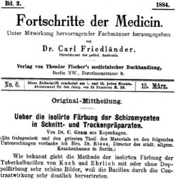
“Ueber die isolirte Faerbung der Schizomyceten in Schnitt- und Trockenpraeparaten”

The Giant’s Shoulders blog carnival is coming up in two days, and I just realized I still haven’t gotten a post up for it yet. So, here it is.

I put up some quick reviews of several classic microbiology-methods papers for the previous edition of this blog carnival, but didn’t actually get around to putting up the one for what is almost certainly the most well-known microbiology technique: “The Gram Stain”. So, this post is about it:

Gram HC: “Ueber die isolirte Faerbung der Schizomyceten in Schnitt- und Trockenpraeparaten”; Fortschritte der Medicin; 1884; vol 2, pp 185-189

That’s “Regarding the Isolational(?) Coloring of Schizomycetes in Cut- [i.e. tissue sections] and Dried Preparations” in “Medical Progress”. The translation hosted by the American Society for Microbiology uses the word “Differential” where I’ve put “Isolational” – which is probably not quite right either but it’ll have to do for now – but I’ll get to that in a moment.

If you’ve ever been exposed to microbiology labwork before, you’ve almost certainly done or at least watched a procedure referred to as a “Gram stain”. In brief, you smear your sample with bacteria on a glass slide and bake it on, then you dump some purple stuff on it, them some brown stuff, then you rinse it briefly with alcohol, then you dump on some pink stuff, and then rinse it in water and look at it under a microscope. Bacteria that stay the original dark purple-blue color of the original purple/brown stuff are considered “Gram Positive”, and those that don’t instead appear the pink color of the last stain, and are considered “Gram Negative”. Many textbook authors and microbiology instructors will breathlessly proclaim that the Gram Stain reveals two “fundamental” categories of bacteria, but I’ll spare you my rant about that.

Properly speaking, this isn’t actually Gram’s stain, as described in his original paper. The modern variations that we’re all taught in microbiology class were developed later, and I believe they are nowadays based mainly on Victor Burke’s 1922 paper on the subject[1].

Regarding the title of the paper: “schizomycete” is what they used to call most kinds of bacteria. “Mycete” meaning “fungus”, as bacteria were assumed to be “plants without chlorophyll” just like molds and mushrooms, and “Schizo-” meaning “split in two”, since bacteria reproduce by splitting into two cells rather than by producing spores like “other” fungi. I say “most” because things like cyanobacteria (“blue-green algae”) or Green Sulfur Bacteria would have been referred to as “Schizophyta” (“fission-plants”). What Gram was originally trying to do wasn’t to differentiate one kind of bacteria from another, either, but to make it easy to tell bacteria from from the nuclei of cells in bacteria-infected tissue.

For that matter, Gram was really metaphorically standing on the shoulders of Koch and Erhlich, as he was building on their technique for staining “tubercle bacteria” – that is, tuberculosis-causing members of the genus Mycobacterium. Gram mentions that you need to stain this type of bacteria for the “usual” 12-24 hours to make this work, incidentally, as opposed to a few minutes for other “schizomycetes”. This suggests that you are expected to have some idea of what you’re going to find before you use the stain, as opposed to the modern implementation which is supposed to tell you something about what kind of bacteria you’re finding.

Still, Gram does report that some bacteria take the stain and some don’t, giving us a preview of the “differential” character of the modern version. He specifically notes typhoid and some causes of bronchial pneumonia fail to hold the stain. Given that Typhoid Fever is caused by a strain of the “Gram-negative” butt-bacter Salmonella enterica, and there are a number of “Gram negative” bacteria as well as “Gram positive” that can cause pneumonia, this makes sense. He also does mention the use of Bismarck Brown R a.k.a. Vesuvine as a counterstain in order to make the nuclei of the infected cells brown in contrast to the dark blue of the infectious bacteria in the tissue.

For much of the century-and-a-quarter since Gram’s publication, the question of why the Gram stain works was thoroughly investigated, and even today I occasionally hear or read assertions to the effect that the Gram Stain isn’t well understood. I disagree with this just as I think its importance to bacterial identification is grossly overblown, and if you want to know why, I have a previous post all about why the Gram stain works and how we know. You may or may not also be interested in an older post regarding whether or not “acid-fast” bacteria like the ones that cause tuberculosis (which don’t stain at all when using the modern version of the Gram stain) are “Gram Positive” or not. As always, if you spot any errors or have any questions, please let me know…

[1] Burke V: “Notes on the Gram Stain with Description of a New Method.” J Bacteriol. 1922 Mar;7(2):159-82.
“A simplified method of staining endospores”

One more for the “classic papers” challenge:

Schaeffer AB, Fulton MD: “A Simplified Method of Staining Endospores”; 1933; Science; 77; pg 194

If you take a microbiology lab, this is the endospore staining technique (or “technic” as they used to spell it) that you’ll practice. This is a nice, simple, one-page paper. Alice B. Schaeffer and co-author Mac Donald Fulton describe a few of the other variations on endospore staining techniques, then describe how they’ve further simplified what they felt was previously the simplest one, described by a Mr. Wirtz in 1908.

“Endospores” are a sort of “escape pod” for certain specific kinds of bacteria. Unlike spores formed by yeasts and molds, these are not reproductive – each bacterium only produces one thick-coated spore, into which it shoves it’s genetic material and a few vital enzymes to get itself going again later when the spore finds itself in favorable conditions.

Since only a few kinds of bacteria produce these endospores, if you see endospores in your unknown bacterial culture it goes a long way towards helping to identify the bacterial species, so having a simple method for staining your bacteria so that endospores are obvious under a microscope is helpful. (Of course, these days most of us would rather just get a 16s rDNA sequence with PCR, but never mind that for now…)

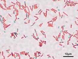
Endospore stain under a microscope (via Wikipedia)Evidently, Wirtz’s original method involved using Osmium Tetraoxide (“osmic acid”) to stick the bacteria to the slide before staining. Not only is that stuff poisonous, it’s also expensive. I found a site selling sealed glass ampoules containing 1 gram each of this stuff for $35.00 each. Schaeffer and Fulton’s method does away with this in favor of much cheaper and easier heat-fixing (just as is done with the Gram stain and others). They use the dye “Malachite Green” for the initial stain, and steam-heat the dye-covered bacterial slide a few times to sort of “cook” the dye into the thick-walled endospores if they are there. Rinsing then washes the dye out of everything but the endospores, and a light red dye (safranin) is added as a counterstain. The end result is that under the microscope you’ll see light-red bacteria. If any of them form endospores, you’ll be able to see them as smaller green dots – sometimes still bulging inside of bacterial cells, sometimes floating around freely having escaped from the now-empty bacterial cell.

The “Schaeffer-Fulton Endospore Stain” is pretty easy to do, though the occasionally messy steambath part can be annoying. The method is pretty resistant to errors, so it’s not too hard to get good results even if you’ve never done it before.

Incidentally, you can buy Malachite Green at many pet stores – it’s still used as a treatment for “ick” (Ichthyophthirius infestation) in tropical fish.

Hmmm…still a couple of hours before it’s not longer May – Perhaps I can throw in one last post before time’s up…

“A small modification of Koch’s plating method.”

Only two more days for the Classic Papers Challenge, so if I’m going to get any more up, I’d better get my butt in gear.

Here’s a nice easy one:

Petri, R. J.:”Eine kleine Modification des Koch’schen Plattenverfahrens.” Centralblatt für Bacteriologie und Parasitenkunde; 1887; Vol. 1, pages 279-280.

The American Society for Microbiology has a translation available online. It’s only about a page-and-a-half of relatively large type – check it out.

There’s a trick we microbiologists use for growing bacteria. You make a solid (but wet) surface that contains whatever nutrients the microbe (bacteria, archaea, yeasts, mold spores…) you’re interested in need, and then you spread a diluted mixture of the microbe on it. The idea is that since the surface is solid the microbes can’t move around too much, and at any spot where a single cell starts initially, a whole pile of that cell and it’s genetically-identical (non-sexually-produced) clone-children will form until it gets big enough to see without a microscope. This cell-pile is called a “colony”, and you can poke or rub it with a sterile object, then stick the object into a sterile nutrient source. The end result is you have a “pure” culture of microbes that are effectively genetically identical. The solid material could be a lot of things – I’ve seen references to using slices of potato – though these days agar-agar gel mixed with nutrients is the preferred substance.

Koch (that is, Robert Koch of “Koch’s Postulates” fame, not Ed Koch the former mayor of New York City) used gelatin (so, hey, here’s another thing you can do with your expired Jell-O®). He apparently used to have a stack of shallow bowls, and had to use a special pouring device to carefully dump the gelatin into each stacked bowl in turn, then cover the works with a bell jar in order to keep stuff from falling into them from the air and contaminating them.

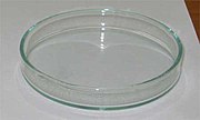
This was kind of a pain to work with, so some clever guy named Julius came up with a modification of this method in 1887, using pairs of shallow dishes, one slightly larger than the other so that it could be turned upside down to use as a lid. Then, you don’t necessarily need the bell jar, and you don’t need to stack them so they’re easier to pour.

Julius Robert Petri’s idea was so useful that we still use it today. Oh, yeah, and they named the dish-and-lid combination after him.

How’s that for a “classic” paper?

Meanwhile, my “Mountain Dew® Wine” project is turning out to be substantially more educational and fascinating than I’d hoped. There seems to be a decent amount of information available on how benzoic acid affects yeasts. I intend to turn that into a post later, but first I’ll try to find at least one more old paper to post before tomorrow is over…