The Gram Stain Post to End All Gram Stain Posts

Gram stain, Gram stain, Gram stain! Bah. I think it’s time Microbiology grew up and moved out of Medicine’s basement.

Sure, the Gram stain[1] has its uses, but the procedure is grossly over-hyped. “[…]the most important stain in microbiology[…]”[2]! “[…]it is almost essential in identifying an unknown bacterium to know first whether it is Gram-positive or Gram-negative.”![3] “The Gram Stain reaction is an especially useful differentiating characteristic.[…]The Gram reaction turns out to be a property of fundamental importance for classifying bacteria phylogenetically as well as taxonomically.”![4] “[…]differentiates bacteria into two fundamental varieties of cells.”![5] “The Key to Microbiology“![6] [emphasis added…]

Bah! Sure, the Gram stain has its uses, but the hype it gets (even 125 years after its invention) is ridiculous. It’s worse than Harry Potter!

You really want to know what the Gram reaction tells you? Really? Okay, here it is:

A “Gram Positive” reaction tells you that your cells have relatively thick and intact cell walls

A “Gram Negative” reaction tells you that they don’t.

That’s it. That’s about all you can reliably infer from the Gram stain.

Previously, I put up a post describing what was my understanding of the conventional view of why the Gram stain works. Today, I’ll give you a much more detailed – and more correct – explanation of why it works as well as what its real significance is to identification of microbes. But first, a brief one-paragraph rant on why I think the Gram stain has such a hold on microbiology teaching.

I blame the fact that microbiology education is still largely in the shadow of medical technology education. When you artificially exclude the 99+% of organisms that aren’t associated with human diseases, the tiny number left do, indeed, seem to largely separate into two phylogenetic categories. Judging by what I’ve encountered thus far, it seems you get a lot of Proteobacteria (especially ?-Proteobacteria, like E.coli), which are “Gram-negative”. You also get a lot of Firmicutes (Bacillus, Streptococcus, Staphylococcus, etc.), and a couple of scattered Actinobacteria (Mycobacterium, for tuberculosis and leprosy, Corynebacterium for diptheria…). Both of these are considered “Gram-positive” (although if you use the standard procedure these days, the Mycobacteria may show no reaction at all). That’s, what, 3 phyla out of about 25 eubacterial and archael phyla? If we throw in Syphilis and Chlamydia, that’s still only 20% or so of the currently recognized prokaryotic phyla. If your microbiology classes assume everybody is training to be a medical technologist or clinical microbiologist, then the Gram stain becomes inflated in importance.

Enough of that – here’s a quick review of how the Gram stain works. Solutions of “Crystal Violet” (a purple dye) and Iodine are applied to cells fixed to a slide, where they soak in and precipitate in the cells. A “decolorizer” (usually ethanol) is applied to see if it will wash this dye precipitate out of the cells. A different, lighter-colored dye (such as safranin) is added so that the cells which DO have their dye washed out can be seen as well. In the end, “Gram positive” cells are a dark purple from the crystal violet/iodine that was not washed away, and “Gram negative” cells are not dark purple. (Usually they are pink, from the safranin, assuming that’s the dye used as the counterstain.)

Note that this does not differentiate cells into “two fundamental types” as is often claimed. You actually get four types: Groups of cells that are normally always “Gram positive”, Groups of cells that are normally always “Gram negative”, Groups of cells that are normally sometimes “Gram positive” and sometimes “Gram negative” (“Indeterminate”, or as I like to call it, “Gram-biguous”), and groups of cells that are normally NEITHER Gram-positive nor Gram-negative, like Mycoplasma, which aren’t dyed at all by the process. Incidentally, phylogenetically speaking, Mycoplasma is one of the “Gram positive” Firmicutes, just like Bacillus and Staphylococcus.

It’s kind of interesting to me that the Gram stain reaction has been such a mystery up until a century after its invention. What is it that makes “Gram positive” cells retain the dye while “Gram negative” ones don’t? Along the way, it seems like nearly every part of the bacterial cell was hypothesized to be the reason for the Gram reaction – lipids, carbohydrates, nucleic acids, “Magnesium ribonucleates”, and so forth. Davies et al, 1983, includes a table listing many of these and referencing historical papers making the claims. The fact that the reaction had something to do with the cell wall seems to go back quite a while, though the “Magnesium ribonucleates” idea doesn’t seem to have been entirely abandoned until the mid-1960’s[7]. It was also hypothesized that the “Gram positive” cells simply absorb more dye and therefore take longer to “decolorize”.

It turns out that “Gram-positive” cells actually don’t, necessarily, take up more dye than Gram negative ones. This was tested by taking a set concentration of bacterial cells and adding them to a set concentration of dye. After letting them soak, the samples were centrifuged to remove the bacteria, and the amount of dye found to be missing from the liquid was taken as the amount absorbed by the cells. They found that some Gram negative cells actually took up more dye than the Gram positives did. So much for that idea.[8]

Even relatively recently, I’ve seen it written that the bacterial cell wall, specifically, is what holds onto the stain, but even that turns out not to be true. Although the cell wall is the structure that seems to be responsible for the Gram reaction, in the late 1950’s it was demonstrated that it was not actually the staining of the cell wall that caused the reaction, but rather the ability of the cell wall to keep the decolorizer out of the cell.[9]

Apparently, the Crystal Violet/Iodine complex itself doesn’t even play a vital role. The complex apparently dissolves again more or less instantly as soon as the decolorizer touches it[10], and it’s even possible to differentiate “Gram positive” and “Gram negative” with simple stains like methylene blue or malachite green, if you’re clever about it[11]. The latter authors set up a clever test with crushed cell material, dye, and paper chromatography. They had the decolorizer soak into the paper, past a spot where dye-soaked cell material from Gram-positive and Gram-negative cells was placed, and watched for obvious differences in the amount of time it took the dye to be carried out by the decolorizer. Incidentally, my quick examination of this paper makes it look like cheaper 100% isopropyl alcohol (“rubbing alcohol”) might be slightly better than the standard 95% ethanol for Gram stains.

– INTERLUDE –

So, here we are at 1970 or so, and we already know that the Gram reaction is entirely based on how well the cell wall structure prevents organic solvents (like ethanol) from soaking into the cell to dissolve the dye complex. Yes, the mystery of why the Gram stain works in normal cells was largely solved by the Nixon era.
A few corners of the mystery remained, though. Why do “old” cultures of “Gram positive” cells often end up staining “Gram negative”, for example? Why do some kinds of cells seem to be sometimes Gram positive and sometimes Gram negative in the same culture? What, exactly, is really happening to the cell, deep down, during the staining process?

In 1983, the Gram Stain made the great technological leap into the 1930’s, when a variation of the technique was devised which allowed the Gram Stain to be observed by electron microscopy[12]. Using a funky platinum compound in place of iodine, the electron microscope reveals exactly where the dye complex is at any particular stage of the Gram stain process. Using this technique, it was possible to see how the decolorizer disrupts the outer membrane of classically-Gram-negative organisms and to see that the decolorizer potentially damages the cell wall and interior membrane, possibly allowing cell material to leak out (or decolorizer to get in and dissolve the dye complex). It was also seen that the dye complex permeates the entire cell, not just the cell wall.[13]

If you’ve been wondering about the sometimes-Gram-positive-sometimes-Gram-negative cells, the same technique was also used to investigate this. As suspected, it turns out that the “old cultures become Gram negative” problem is due to the cell walls breaking down as the culture ages. Bacteria are continuously, simultaneously, building up and tearing down their cell walls, in order to be able to grow and divide. As nutrients run out, the bacteria run out of material to rebuild cell walls, while the cell-wall degrading enzymes keep on chugging. Breaks in the cell wall occur, and through these breaks the decolorizer can get in and rapidly dissolve the dye. Actinobacteria can have a similar problem, but rather than only being in “old” cultures, apparently weaknesses appear briefly during cell division, and if a particular cell happens to be at this stage of growth when you stick it on a slide, heat-fix, and Gram stain it, the weakness at the septum where the division is occuring can crack and allow the decolorizer in, resulting in a “Gram negative” response even while surrounding cells of the same kind might still be “Gram positive”.[14]

This brings us to archaea and some eukaryotes (i.e. yeasts). Yeasts stain “Gram positive” normally. Although their cell walls are completely different chemically than bacterial cell walls, they are quite thick (microbially speaking). Poor, neglected Archaea seem to be all over the place in terms of Gram reaction. Since their Gram reaction doesn’t tend to correlate to any particular phylogenetic grouping[15], it seems nobody really pays much attention to their Gram stain reaction. On the other hand, and on the subject of “Gram-biguity”, I thought the investigation of Methanospirillum hungatei[16] was interesting. M.hungatei is an archaen that grows in chains. When Gram-stained, the cells on the ends of the chains are “Gram positive”, while the others have no Gram reaction at all. It turns out that the chains are covered by a sheath, and the only contact with the outside world is through thick “plugs” in the cells at the ends of the chains. These “plugs” act like thick cell walls, allowing the Gram stain dye material to soak in but excluding the decolorizer, while the sheath keeps the rest of the cells from soaking up any stain at all.

There you have it – a relatively detailed history and explanation for the Gram stain, and you didn’t even have to get through some obnoxious paywall to read it. Aren’t you lucky?

Comments, suggestions, and corrections, as always, are welcome.

[1] Gram, HC.”Ueber die isolirte Faerbung der Schizomyceten in Schnitt-und Trockenpraeparaten.” Fortschitte der Medicin. 1884 Vol. 2, pp 185-189.

[2] Popescu A, Doyle RJ. “The Gram stain after more than a century.” Biotech Histochem. 1996 May;71(3):145-51.

[3] Brock TD, Madigan MT, Martinko JM, Parker J. “Biology of Microorganisms (7th Edition).” 1994. Prentice Hall, Englewood Cliffs, NJ pg. 46

[4] ibid, pg. 715

[5] Beveridge TJ.”Use of the gram stain in microbiology.” Biotech Histochem. 2001 May;76(3):111-8.

[6] McClelland, Rosemary. “Gram’s stain: The key to microbiology – isolate identification method – Tutorial” Retrieved 20070810 from http://findarticles.com/p/articles/mi_m3230/is_4_33/ai_74268506/print

[7] Normore WM, Umbreit WW.”Ribonucleates and the Gram stain.” J Bacteriol. 1965 Nov;90(5):1500.

[8] BARTHOLOMEW JW, FINKELSTEIN H:”CRYSTAL VIOLET BINDING CAPACITY AND THE GRAM REACTION OF BACTERIAL CELLS.” J Bacteriol. 1954 Jun;67(6):689-91.

[9] BARTHOLOMEW JW, FINKELSTEIN H.”Relationship of cell wall staining to gram differentiation.” J Bacteriol. 1958 Jan;75(1):77-84.

[10] LAMANNA C, MALLETTE MF. “CHROMATOGRAPHIC ANALYSIS OF THE STATE OF ASSOCIATION OF THE DYE-IODINE COMPLEX IN DECOLORIZATION SOLVENTS OF THE GRAM STAIN.” J Bacteriol. 1964 Apr;87:965-6.

[11] Bartholomew JW, Cromwell T, Gan R.”Analysis of the Mechanism of Gram Differentiation by Use of a Filter-Paper
Chromatographic Technique.” J Bacteriol. 1965 Sep;90(3):766-77.

[12] Davies JA, Anderson GK, Beveridge TJ, Clark HC.”Chemical mechanism of the Gram stain and synthesis of a new electron-opaque marker for electron microscopy which replaces the iodine mordant of the stain.” J Bacteriol. 1983 Nov;156(2):837-45.

[13] Beveridge TJ, Davies JA.”Cellular responses of Bacillus subtilis and Escherichia coli to the Gram stain.” J Bacteriol. 1983 Nov;156(2):846-58.

[14] Beveridge TJ. “Mechanism of Gram Variability in Select Bacteria.” J Bacteriol. 1990 Mar;172(3):1609-20.

[15] Beveridge TJ, Schultze-Lam S. “The response of selected members of the archaea to the gram stain.” Microbiology. 1996 Oct;142 ( Pt 10):2887-95. (Abstract)

[16] Beveridge TJ, Sprott GD, Whippey P. “Ultrastructure, inferred porosity, and gram-staining character of Methanospirillum hungatei filament termini describe a unique cell permeability for this archaeobacterium.” J Bacteriol. 1991 Jan;173(1):130-40.

Simplify, Simplify…

DNA seems to have two main threats to its well-being once it’s extracted and purified.

  • Nucleases
  • Spontaneous Hydrolysis by water

Nucleases are the big one that everyone seems to mention. The seem to be fairly sturdy enzymes, and they’re everywhere (including fingertips – hence the need to wear gloves whenever you get near DNA samples…), and they “eat” DNA rapidly. Theoretically, you can destroy the enzymes with enough heat, but you still need to worry about them getting in every time you pop open your sample to get some out.

Apparently, DNA even in pure water can tend to slowly fall apart spontaneously. It doesn’t happen very fast, but bit by bit, it can undo the links between the individual nucleotides.

A common way to try to deal with nucleases is to add EDTA to the solution. Nucleases need magnesium ions dissolved in the water to do their job, and EDTA tightly binds to magnesium (and calcium). The idea is to “use up” any stray magnesium ions in the solution so that even if nucleases get in, they’re inactive because they have no magnesium available. That’s why you see EDTA in the recipes for so many DNA-related solutions. Of course – EDTA doesn’t permanently bind up all the magnesium – there’s always a tiny fraction that stays in the solution. So, although EDTA can drastically slow down any nucleases, it won’t actually stop them.

There are also some interesting chemicals which can be added to destroy all proteins (including nuclease enzymes). Guanidine Thiocyanate is one rather nasty chemical that does this. 2-mercaptoethanol is another. Various other detergents like CTAB may also denature any proteins. Since they don’t harm the DNA in the process, you could keep the DNA sample dissolved in a solution with these chemicals…but then you can’t do PCR with the sample as it is, since the protein-denaturing chemicals will also destroy any enzymes that you WANT, like DNA Polymerase, when you try to mix it into your reaction.

I think the latter option will be great for collecting field samples (in fact, it’s papers specifically on the subject of preserving samples in the field with CTAB and Guanidine Thiocyanate based solutions that I’m adapting from), but isn’t going to be real useful once I’ve got my DNA relatively purified. What to do, what to do…

Actually, I think the answer’s simpler than I originally expected. I’ll just dry the purified DNA out. No water – no hydrolysis…and no nuclease activity, either.

I could actually just leave it as a dried pellet in the bottom of a microcentrifuge tube, but that leaves the problem of taking only a little bit of it for processing rather than taking the whole thing, and I want to avoid reconstituting it and re-drying it repeatedly. I think a variation of the “dry the DNA on a piece of paper” process will be in order – then I can just cut off a small strip of the paper to get a portion of the DNA. It appears that you can actually dunk the DNA-impregnated bit of paper right into whatever solution you’re using (like a buffered polymerase-and-primers solution for PCR) and go for it.

Among the several references I found on this, here are two:
Kawai J, Hayashizaki Y: “DNA Book”; Genome Res. 2003 13: 1488-1495
Burgoyne LA: U.S. Patent #5496562 “Solid medium and method for DNA storage” (1996); U.S. Patent and Trademark Office, Washington D.C.

Okay then…

My summer classes are finally over. Got an “A” in immunology (go, me). Now I just need to make sure everything’s done next semester. I’ve already signed up for the last two Underwater-Basket-Weaving-type “General education” classes required at this college: Intro to Philosophy and “History of Western Art”. I also went ahead and signed up for Environmental Chemistry, too – it’s not required, but it’s one of the last “not required but useful if I have time for it” classes on my list.

Meanwhile – is it just me, or is DNA some obnoxiously fragile stuff when you don’t want it to be? Sure, leave a few flakes of skin or hair follicles at a crime scene and they’ll nail you weeks or months later, but try to “gel purify” some DNA and it just falls apart…

The samples from my last post, about the colony PCR of my Lactic Acid beer-bacteria, I cut the bright bands of presumably-16s rDNA out of the gel and ran them through one of those canned “gel purification kit” processes. Then I froze them until I had a chance to finish my classes and play with them.

Yes, I was wearing gloves. No, I didn’t lick the gel. I think I must have looked at them too closely or something and they just disintegrated out of spite. In any case, my attempt at a restriction enzyme digest turned up NOTHING (other than the “ladder” lanes) on the gel.

I’m beginning to really distrust canned kits. On the upside, that means I get to learn some more in the process of developing my own replacement protocols.

I will probably try re-amplifying DNA from the frozen samples and see if there’s anything at all left in there that can be saved. Otherwise, I’ll also check and see if the plates I made a few days ago still grew okay.

In other news – I’m toying with the idea of literally begging for my own microscope and home-microbiology lab equipment. As in, actually putting on a lab coat, taking an old hat, and sitting outside of scientific meetings and such with a cardboard sign saying “want my own microscope – please help”. Of course, I’d have to report any donations as “income” for tax purposes – I doubt they’d let me form a 501(c)(3) corporation dedicated to just buying me toystools for my own microbiological amusement.

I haven’t decided, but it’s under active consideration. It’d make for some interesting blogging (and I promise in return that I’d account on the blog for any money donated, and blog all uses of the equipment under Creative Commons terms so everyone can use it). It’d presumably take a while for this to get anywhere if it ever did – it seems it’ll cost about $400-$500 just for a (good) basic light microscope, plus another few hundred for a darkfield condenser and related upgrades. Plus, of course, me wanting to build some LED-based lighting for fluorescence microscopy ($500 canned commercial upgrade? Bah!). Incidentally, it seems Green Fluorescent Protein fluoresces best right around the wavelength of a typical, inexpensive, off-the-shelf ultraviolet LED…

And then of course I need a pressure cooker and one or more incubator setups and some petri dishes and trips to the grocery store for growth media and staining supplies and slides and… well, anyway, as much stuff as I can arrange to get. But the microscope is the one component that is unavoidably expensive.

Oh, yeah, and some space to keep cheese and beer culture organisms and such for later use…

Comments, anyone? Suggestions?

I, for one, WELCOME our new radiation-eating fungal overlords…

Though I am getting a little annoyed at the breathless prose about how it’s “like photosynthesis” and might be a way to sustain astronauts during long space flights and so on.

The story’s about a fungus they found growing (thriving, even) inside the reactor at Chernobyl, despite all the radiation the fungus is exposed to in there.

What the original paper – which you can find here from PubMed Central (and where you can find what the study actually shows, rather than the somewhat lower-content hype found in most news reports on it) – seems to show based on my hasty undergraduate-level reading is that the fungi do grow faster when exposed to “ionizing radiation”, and that it appears to be due to melanin in the plant getting energy from the radiation (and passing that energy on to the fungus to use for growth).

This is actually pretty spiffy, but really – so far – doesn’t look like “photosynthesis” at all. They’re not testing for any kind of carbon fixation, and I’m guessing that if there is any carbon fixation going on, that it doesn’t generate oxygen in the process. It also seems unlikely to me that even then, the fungus can grow autotrophically. This would seem to drastically reduce the possibility of this stuff ever being Purina® Astronaut Chow – you’d still need some other way to get the carbon dioxide out of the Astronaut’s air and put oxygen back in it. If you’re going to do that, you might as well just use plants (or cyanobacteria) and eat THEM.

Still, the implication that you could adapt some melanin-producing fungus to absorb “radiation” and turn it into useful materials of some kind is spiffy, even if it’s not going to allow us to turn nuclear fission plants and spent nuclear fuel depots into fungus-powered anti-global-warming-gas powerhouses.

One thing’s bugging me, though. I obviously don’t have enough understanding of how “ionizing radiation” behaves at a biochemical level, since I’m wondering if it’s proper for everyone to treat “radiation” (both from flying electrons and from high-energy light) as some sort of generic substance, whose only useful attribute is how much energy it has.

As far as I know, most of the “radiation” that the fungus inside the Chernobyl reactor is getting is Gamma-radiation – basically high-energy light (one step above “X-rays”, two steps above sunburn-causing Ultraviolet light). What the researchers are hitting their test-subjects with looks like it’s mainly “Beta”-radiation (which is to say – electrons)*. In both cases it’s “ionizing” radiation, which is to say (more or less) that the radiation knocks electrons off of atoms that it runs into in both cases, and in the ideal “spherical horse” world of a Physicist, the same amount of energy is going to knock the same amount of electrons loose from various molecules and therefore have the same effect, right?

Except I’m having trouble convincing myself that’s a valid assumption here. The results seem to show that exposure to radiation is somehow resulting in the melanin in the fungus being able to “reduce” a chemical (changing “NAD+” into “NADH”) that can potentially in turn dump electrons into the beginning of the Electron Transport Chain to in turn provide biological energy in the form of ATP…

Can one reasonably assume that the mechanism by which this happens would be the same regardless of the form of ionizing radiation? The big deal with melanin seems to be that it absorbs a wide range of light wavelengths (which is why it looks black to dark-brown, and why it protects skin from Ultraviolet radiation…) which implies that absorbing the gamma radiation is where the energy is coming from that makes the fungus thrive in the Chernobyl reactor building. I guess I’m just having trouble picturing how a much more massive, slower-moving electron could have precisely the same effect as a virtually massless, much faster photon. (Yes, I know that beta and gamma radiation are said to have the same amount of “effect” on living tissue per unit of energy…)

Is it possible that the melanin is directly “capturing” the beta particles (electrons), while gamma radiation is kicking electrons off of something ELSE, and melanin is then only indirectly taking up those? For that matter, is it possible that in both cases it’s just something silly like the radiation inducing hydrolysis of water, and it’s just hydrogen gas supplying the reducing power? Thinking about this is making me feel dumb – can anyone reading this explain what I’m missing here?…

I suppose I could just cheat and ask someone in the biology department. We’ve GOT a professor who ought to know – her research has specifically focussed on zapping prokaryotes with “ionizing radiation” (electrons from the college’s linear accelerator)…But that would rob my dear readers of the chance to participate here…

* – okay, it’s probably even more complicated than that. If I understand what the paper is describing and what my Minister Of Funky Physics Knowledge showed me, the source of the “ionizing radiation” for the experiments is radioactive Tungsten(W) and Rhenium (Re) (A “188Re/188W Isotope Generator”). W-188 gives off beta particles when it decays to Re-188. But Re-188 can go through some sort of funky subatomic rearrangement before it decays so that it can EITHER give off beta particles OR gamma-rays as it decays down to stable Osmium-188. I have no idea what the proportion between beta and gamma is at that step (the “conversion efficiency”) so it’s possible there’s enough gamma radiation coming out to do something, regardless of what the beta particles are doing. (The experiment doesn’t do any comparisons with “pure” gamma radiation, which I imagine is not simple to arrange…). So now I’m even MORE confused. Thanks, physics. Thanks a lot.

Colony PCR – because DNA extraction protocols suck.

If you’ve got a culture of a single type of bacteria and you want to identify it, the standard method is to figure out the sequence of one particular gene, the 16s rDNA gene. That is – it’s the gene which encodes the a piece of RNA that gets used by the ribosome in part of the process of “reading” which amino acids to link together to make a particular protein. This is something that every prokaryote known has, and parts of it are conserved, so they’re similar enough to compare, while other parts can vary a lot, providing enough “difference” to tell different organisms apart.

To figure out the sequence, you use PCR to “amplify” this particular gene, making lots of copies of it so that the sequencing machine can clearly see the signal from each part of the sequence. And before you can do that, you have to get the DNA out of the cell relatively intact.

That part can be a pain. There are lots of different ways people have come up with (and made special canned “kits” out of) – you can use chemicals to try to dissolve the cells and let all their guts (including the DNA) out, you can try to mash them up with tiny glass beads in a “bead-beating” machine, you can stick them in a blender, you can even just boil them for a while…then usually you go through several steps of centrifuging and mixing with different chemicals and then centrifuging again until you’ve hopefully finally got the DNA out and gotten rid of most of the other cell bits. And, hopefully, you haven’t accidentally chopped up the DNA too much to use in the process.

Fortunately, there’s a trick you can sometimes use, referred to as “Colony PCR”. In it, you literally just touch the top of your colony of cells and shake them off directly into the PCR tube. Then you just include an extra 5-10 minutes of 95°C heating to hopefully cook open enough of the cells to release DNA (and cook the cell’s enzymes to death so they don’t degrade the DNA and interfere with the PCR).

Not real reliable if you’re trying to do anything quantitative, like trying to figure out how many copies of a gene are in each cell, or trying to get an accurate estimate of how many cells of one type or another are in a mixed culture, but if you just need as much of a particular bit of DNA as you can get – such as for sequencing – a lot of people use this.

I just tried it on my Lambic isolates. Two of the 8 bacterial cultures worked beautifully. I’m pretty sure the problem with the other 6 was just the sheer amount of bacteria I ended up adding to the reaction – too much seems to “swamp” the PCR process and keep it from working. I’ll try it again this week. But it does seem to indicate that it works, at least.

Tasty Acids

The story so far – I’ve got 8 live bacterial cultures (and two yeasts) obtained from a bottle of Peach Lambic, imported from Belgium. I strongly suspect that 6 of the 8 are Pediococcus species, and the other two are in the Lactobacillus genus. It is also possible that some of them might turn out to Leuconostoc or some other genus, but I suspect them all to be in the Order Lactobacillales somewhere, anyway.

Hopefully I’ll be able to get good, definitive sequence data from the bacterial isolates later this week.

Lactic acid seems to be the predominant acid in Lambic ales, produced by the various bacteria which break down the sugars in the beer and spew out lactic acid as a waste product.

Pediococcus also shows up in wines, where it’s associated with “malolactic fermentation” – where it converts the harsher malic acid into the more mellow-tasting lactic acid.

Thinking about this led me to think about the other distinctive acids found in foods. Here’s a listing (in no particular order) of some, with foods associated with their distinct flavors:

  • Lactic acid = “Yogurt” acid (and Sour Cream.  And many types of pickles.)
  • Malic acid = “Apple” acid (“Green Apple” flavor)
  • Tartaric acid = “Grape” acid (Verjuice and “Grape flavor”)
  • Acetic acid = “Vinegar” acid
  • Citric acid = “Lemon/Lime” acid (or “Pixy Stix®” flavor)
  • Propionic acid = Swiss cheese acid

In other news, I need a real microscope of my own.

E.coli – the “Microsoft” of the biotech world?

…by which I mean, it’s not always the best tool for the job, but everyone insists on always using it anyway, and has a variety of excuses for doing so…

Honestly – I’m trying to set up a clone library of 16s rDNA sequences using this kit. Never mind which kit it is – it actually does seem to work. I was just struck by the amount of hassle involved in shipping and storing the kit and it’s supply of “competent cells”.

When you get them, take them out of the dry-ice they’re shipped in and put them in the -80°C freezer immediately or they’ll die! Only thaw them carefully just before you use them, and do it on ice or they’ll die! Don’t heat-shock them for more than exactly 30 seconds or they’ll die! Once you’ve got them growing, you have to keep moving them to fresh selective media frequently or they’ll die! Or, you can carefully place them in the -80°C freezer…or they’ll die! Don’t look directly at them or they’ll die! (Do Not Taunt HappyFunCell!…)…

Seriously, running those gigantic -80° freezers can’t be cheap. Wouldn’t it be more convenient if you could grow up your transformant as an ordinary culture and just add your DNA samples and some kind of inducer chemical to make them take it up? Surely there must be some other organism that might be made to work like that.

Actually, it seems a number of the “Gram-positive” (firmicutes) organisms can enter a state of “natural competence”, where they naturally take up double-stranded DNA molecules from the environment. Bacillus subtilis is one. I’ve even seen references to “natural-competence” based protocols for transforming B.subtilis (or other Bacillus species, presumably) but it only seems to be in an out-of-print, $400 book.

Wouldn’t that be more convenient (using B.subtilis that is, not the $400 book)? Plus, when you wanted to store your transformed culture for later use, you could just heat the culture up to, what, about 55°C for 15 minutes or so (as I recall) then let it dry. The spores will contain whatever “bonus” plasmid DNA you added (if spores didn’t keep plasmids, then anthrax wouldn’t be such a danger…) and will last practically forever at room temperature. Mix the spores with some dried nutrient powder and seal them in a foil packet. Instant transformants, just add water!

But NOOOOO…..”But, everybody else uses E.coli, so I have to.” “They only make ‘BogoGen SuperMiniUltraKlone Kit 2000’ with E.coli, and we have to use that!” “But, nobody knows that other stuff, but everybody’s already familiar with E.coli!” “I’m a BogoGen Certified E.Coli Engineer, and I say everything else is just a toy and doesn’t work!” “All the books and stuff are about E.coli…”

Bah! Pathetic excuses. Anybody got a huge wad of venture capital to throw at me? The more I think about this, the more I think ‘untapped niche’…Heck, the electricity savings on not having to run a -80°C freezer constantly alone ought to qualify for a good “Fight Global Warming – Say ‘No!’ to E.coli!” marketing campaign…

Bonus perk: All the natto you can eat…

More Lambic pictures

Ah, that’s better – a more traditional heat-fix/simple stain (using Methylene blue) shows my yeast isolates better:

Sally the maybe-Brettanomyces-type yeast
(“Sally”, a yeast that I suspect is a Brettanomyces-type yeast.)

Sam the...Saccharomyces-type yeast?
(“Sam” now looks awfully small…but more experienced observers than I am said that it could actually be a Saccharomyces-type yeast.)

Lucy the possibly-Pediococcus
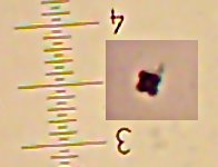
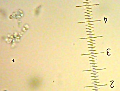
I also got two more Coccoid-Cluster-type Gram-positive bacterial isolates. The look pretty much the same under the microscope, though one had gooey wet, slightly larger colonies than the other’s smaller, hard-lump colonies. I see another one of those
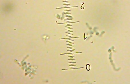
 tetrads in the hard-lump-colony microscope image.

All told, I now have 10 isolates to check out. I’ve been given the go-ahead to try sequencing on the 8 bacterial isolates so hopefully I’ll be able to get a clear identity for Fred, Sid, Lisa, Lucy, BillyBob, JimBob, BettySue, and MarySue. Sally and Sam will have to wait for now, though I’m looking into ways to characterize them, too.

“Live and active cultures” – of beer.

I’ve got a project going to isolate as many yeasts and bacteria as I can from the dregs of a bottle of relatively-famous-brand Lambic ale.

So far, I’ve got at LEAST 3 different types of bacteria and two different yeasts – all of which I suspect are “intentional” – that is, the bacteria are probably lactic-acid bacteria (Lactobacillus, Pediococcus, etc.) which are expected to grow there, and the yeasts I believe to be a Brettanomyces-type yeast and a Saccharomyces yeast (based purely on what I expect to find and the small amount of microscopy that I’ve been able to do so far.)

I have at least one and maybe two different “Gram-positive” rod cultures which I believe to probably be Lactobacillus species. I have several isolates of generic “clusters of Gram-positive coccoids” of which there are at least two different types (which look more or less identical in the microscope, but one of which seems to generate acid while eating mannitol and one that doesn’t).

I have so far named three isolates from Sabouraud agar: Sally, Sid, and Sam.

Sally the Yeast
Sally, the maybe-Brettanomyces-type yeast – 400X magnification (Lactophenol Cotton Blue stain)

Sam the Yeast
Sam, the maybe-Saccharomyces-type yeast – 400X magnification (Lactophenol Cotton Blue stain.)

Sid the [lacto?]bacillus-type-thing
Sid, presumably a Lactobacillus-type bacteria – 1000X magnification (Gram stain)

I’ve also collected four isolates (which may actually just be two different organisms) from an initial inoculation on MSA – BillyBob, JimBob, BettySue, and MarySue. MarySue is the one that seems to be “fermenting” the mannitol.

BillyBob, maybe a Pediococcus?
This is BillyBob (I clipped part of the image and moved it closer to the little “ruler”). The others look essentially the same when Gram-stained.

I’ve also got a bacillus-type (presumably Lactobacillus) critter that showed up on an initial BHI which may or may not be the same as Sid, and I got two more BillyBob/MarySue type colonies on another MRS agar plate.

Interestingly, when I did the original inoculations, it’s the ones that I added the LEAST amount of beer sediment to (20?l) that seems to get the growth – higher amounts may just add so much sugary solution (this stuff is quite sweet) that it inhibits growth.I really hope I can arrange to do molecular analysis (specifically, 16s rDNA sequences) on at least the bacteria, if not the yeast as well. I’d really like to get good identification of these. Assuming they’re real Lambic organisms, they’re probably already in the databases somewhere and should be readily identifiable – assuming someone will let me use up some supplies.

Beer cures flesh-eating bacteria, Staph, Strep, and Anthrax!*

* – These statements have not been evaluated by the Food and Drug Administration. Beer is not intended to diagnose, treat, cure, or prevent any disease, except for maybe hypobeeremia.

No, the title isn’t really true, exactly. However, it does appear to be true that a major component of modern beer – Hops (Humulus lupulus) flowers, really does appear to inhibit “Gram-positive” (Phylum firmicutes) bacteria.


The plates in the picture, clockwise from the upper-left, are inoculated with Bacillus subtilis, Escherichia coli, Pseudomonas aeruginosa (note the green pigment), and Staphylococcus aureus. ON the plates are 5 sterilized paper disks, each soaked with an extract of (again, clockwise from upper-left) Coriander, Hops flowers [Tettnanger], Cassia oil, Clove buds, and Ground Ginger root.

Except for the Oil of Cassia (“Cinnamon oil”), I took 2.5g of each ingredient, boiled it for 15 minutes in distilled water, soaked sterile paper disks in the water, then stuck the disks on top of plates inoculated with the bacteria in question. The cassia oil is about 10?l of the pure, full-strength oil as a sort of “positive control”. At that extreme concentration, it seemed to keep everything away.
The results are even more dramatic than I expected. For one thing, I expected at least some inhibition by the clove extract. The water was the color of a moderately strong tea and smelled strongly of clove, so I would have expected to have enough for some effect…but, no, it was just too feeble. (Had I used pure eugenol, I’d have probably seen the same effect as with the “cinnamon” oil.) Compared to the rest, a mere 15 minutes of boiling a comparatively mild variety of hops flower seems to very effectively prevent growth of certain types of bacteria – which would presumably include the varieties mentioned in the title of this post.

Hops skin-lotion to appear at hugely inflated prices on health-food-store shelves in 3…2…1…

Incidentally, if it does, I wouldn’t use it. “Gram positive” bacteria make up a substantial portion of the “normal flora” of healthy skin. Killing them off might easily leave room for other bacteria to take over and cause problems.

It does make me wonder about other possible uses of this effect, but I’ll save that for another time.

I’ll close by pointing out how useless allegedly “anti-bacterial” spices seem to be by comparison. Kind of puts the whole ridiculous notion of medieval cooks using spices to inhibit spoilage or to treat “rotten” food in its place, I’d say. It also implies that hops isn’t going to prevent “spoilage” of beer by itself, given that (for example) vinegar bacteria aren’t “gram-positive” types, nor are all the lovely ?-proteobacterial butt-bacter organisms like E.coli going to be affected…at least not by the hops. More experimentation to be performed at some later date.

This is just a simple experiment on the side of the main one I’m performing, where I attempt to isolate as many different viable organisms from a bottle of famous-brand Belgian Lambic ale as I can, hopefully for use in other foods (sourdough? Yogurt? And, of course, beer…) later.