Environmental Chemistry Field Trip – Day 1, part 3

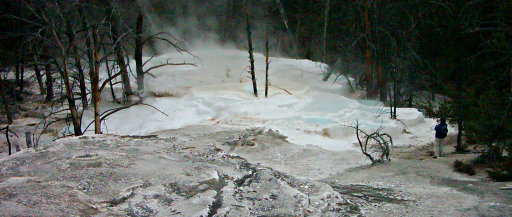
Overview of Narrow Gauge Spring
Our final destination of the day was Narrow Gauge Spring, which is on the backside of the Mammoth Terraces area. Apparently, there’s only one other place in the entire world – somewhere in China – that has exactly the same kind of conditions as this place.

The process of making this kind of formation requires rainwater, healthy microbe-supporting soil, limestone, and heat. It goes something like this: rainwater seeps down through the soil, where lots of healthy microbial activity uses up the oxygen in it and excretes plenty of extra carbon dioxide into it, making it more acidic. The water sinks into the ground and runs into the limestone, which is Calcium Carbonate (CaCO3). Calcium Carbonate doesn’t dissolve well in plain water at all, but there are two things that make it dissolve better: acid and heat. The heat from the magma under the park and the acidity of the water combine to dissolve a whole lot of the limestone. Then, somewhere, the heated water gets forced back up to the surface through a crack.

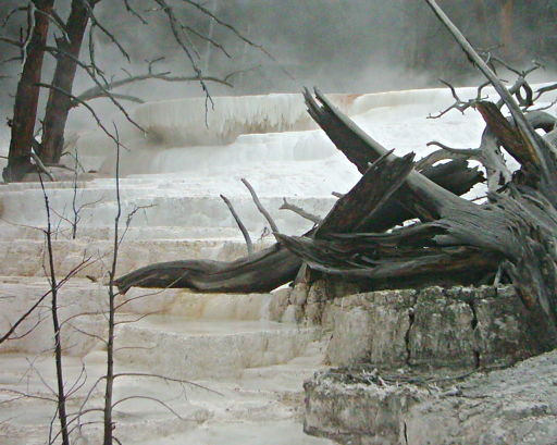
Where the water comes back in contact with the air, it can let off the extra carbon dioxide and heat. This doesn’t happen very fast in a deep pool, since this can only happen in a thin area near the top. Where the water overflows, though, it’s very shallow, and the carbon dioxide and heat can escape very quickly into the air. This makes the water suddenly become less acidic and less hot, and all that extra calcium carbonate can no longer stay dissolved. It crystallizes, making a hard calcium carbonate “shell” along the edge of the pool. The edge can end up growing some much over time that it forms an overhang with stalactite-like formations underneath it:

Another view of Narrow Gauge Spring

You can just make out an overhanging area in the upper-left of the photograph.

It was fun taking measurements of the water here. Water freshly removed from a pool initially showed up off the scale on our “Total Dissolved Solids” meters, but if you waited a few seconds the reading would drop down to where the meters could read it, and keep falling. Out of the pool, the water was cooling off quickly enough that the extra dissolved Calcium Carbonate was un-dissolving out of the water in tiny bits even as we stood there.

The water appeared to be about 56°C at the top of the pool where it was initially emerging. If you want an idea of not only that I am a nerd but what kind of nerd I am, I will mention that I think of this as “stewpot temperature”, and often wonder if there is any useful or tasty effects to be discovered in the microbial processes done by thermophilic microbes that live in these conditions. I’ll find out one of these days…

Oh, and a couple of bits of trivia about the Apollinaris Spring area from a couple of posts ago. Firstly, it was apparently named after a spring in Germany with the same name. Secondly, we briefly discussed the chemistry of carbon dioxide in water in class this week, and it turns out that the pH of 5.9 that Apollinaris Spring has is probably more basic than plain distilled water would be.

Now, anyone who’s had basic chemistry is probably a little baffled by this – after all, isn’t a pH of 7 that of pure water by definition? The answer is yes, but we’re not talking about pure water, we’re talking about water exposed to the air, where carbon dioxide can dissolve into the water. Working through the mathematics involved showed that distilled water should end up with a pH of about 5.6-5.7, at least at “standard temperature and pressure” (roughly sea-level air pressure and a temperature of around 72°F.). I have a suspicion as to why the Apollinaris Spring water seems less acidic than I might have expected, though.

They actually took our Apollinaris Spring water and ran it through an analytical instrument of some kind (I wasn’t there for it, but the description of the results made it sound like it was a “liquid chromatography” type of device). They found NO nitrates or nitrites in it. Since we’re talking about spring water percolating through healthy soil, I would have expected some nitrogen. I noticed, though, that although they checked for nitrite and nitrate, they didn’t check for reduced nitrogen – that is, ammonia.

I managed to score a tiny vial of the water during lab last Wednesday. When I get a chance to hit the pet store for some ammonia testing supplies, I’ll check that. If it’s there, it might explain the possibly slightly higher than expected pH. Similar to what happens to carbon dioxide and water, when ammonia (NH3) is dissolved into water(H2O), there tends to be some recombination of the atoms to make “ammonium hydroxide” (NH4OH), which is basic.

I don’t know if that’s what’s going on, but I intend to check.

There’s one more post worth of Field Trip stuff, and then I’ll be back onto other topics. Here’s a hint of what might come up, though: can anybody tell me what the effective pore size of pectin and cornstarch gels might be?…

Environmental Chemistry Field Trip – Day 1, part 2

Our next stop was Appolinaris Spring, which seems to be an uncommon thing in Yellowstone National Park: ordinary springwater. No sulfuric acid, no steam, just plain old water that sinks into the ground and then comes back up later. For most of the park’s history, it seems like this used to be a popular place to stop to get a drink of water.

water emerging from small copper pipes
Although the signs around the spring now all suggest that you really shouldn’t drink it, at least not without filtering it first, I’m kind of kicking myself now for not having tasted it. Perhaps I’ll have to go back on my own time and try it.

Our on-site tests showed a pH of 5.9 (slightly acidic: milk is normally around 6.8 or so, Root Beer somewhere around a more acidic 4.0, cola beverages around 3.0, for reference…), relatively low TDS of about 100ppm, coming out of the ground cool (about 7°C, or 43°F), with very little dissolved Oxygen (about 6.0ppm) and faintly carbonated (300ppm CO2). It reportedly didn’t taste too good, but having foolishly missed out on tasting it, I don’t know why.

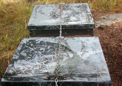
There were hints that perhaps contamination from surface water – like rain trickling through bison poo – but quite some time ago they sealed the spring up to protect it from that kind of thing. This is the actual spring now:

Appolinaris Spring is a concrete box in the ground with locked metal tops...
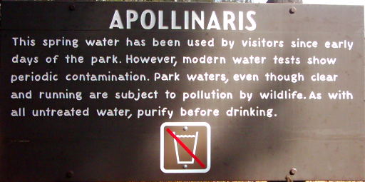
Even so, the signs still try to discourage people from drinking the water coming from the pipes that lead out of the spring, which I take to be the park service covering themselves just in case someone claims to get sick from it. (“Hey, we TOLD you not to drink it!”).

Appolinaris: This spring water has been used by visitors since early days of the park.  However modern water tests show periodic contamination.  Park waters, even though clear and running are subject to pollution by wildlife.  As with all untreated water, purify before drinking.
Periodic pollution by wildlife? What the…

The northern end of a south-bound bisonOh, right. Natural bottled-spring-water flavor. Hey, it’s natural, it’s got to be good for you, right?

And to end this post on a complete and totally baffling non-sequitur: the student lounge I’m sitting in right now has a television constantly tuned to some cheesy mass-media channel. Today it’s “E!®”. I overheard something on it just now that made me sit up and take notice: Evidently “Leprechaun” made a profit. Wow.

One never knows what kind of amazing things one might learn at college…

The Unbearable Limeness of Being

I awaken. Am I alive? The temperature is neither extremely hot nor extremely cold, so I’m apparently not in some punishment-afterlife. And there’s no beer volcano or stripper-factory, so this obviously isn’t heaven. On the other hand, I am experiencing the usual persistent discomfort involved with waking up early in the morning. On the assumption that Catholic “purgatory” would be more dull, I will assume I am still alive, and had better get up and get to class.

Since my previous experiment, I have obviously had to revise my original hypothesis. Since the last caused me no ill effects, I had to abandon the notion that expired gelatin products become a deadly poison. Instead, as I consume this batch of official, non-sugarless Jell-O®-brand Gelatin (Lime flavored), I operate on a new hypothesis:

“Expired instant gelatin products from intact packaging will not harm me if I eat it.”

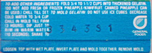
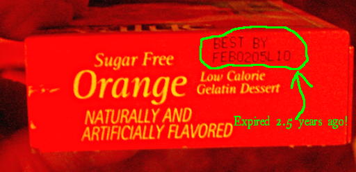
My precious stock of expired JellO® is depleted by one more box, the packet ripped from its cardboard sarcophagus, the contents prepared according to the standard instructions, and consumed hastily last night (the animation from the previous post is the actual container of prepared Lime JellO® made from digital photographs taken between helpings.). You can see the old-style date code on the box. According to Carolyn Wyman’s “JELL-O: A Biography”, the code indicates that it was packaged in 2003 (the “3” at the beginning of the code), on the 343rd day of the year, in the San Leandro (California) packaging facility. Although there is no official “expiration date” shown, given the “expected shelf life” of 24 months, this package is approximately 2 years out of date. And I ate it. I appear to have suffered no ill effects. Not even a decent sugar-rush: the entire box contains 320 calories, barely equivalent to a package of Twinkies®. The flavor even appeared to be perfectly normal. Mmmmmm, Lime JellO…

When I took it out to eat it, I did spot a beautiful if alarming sight, though:

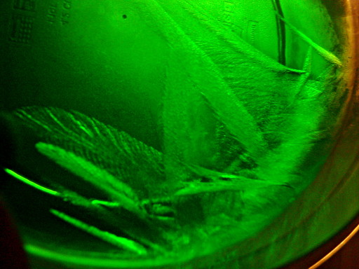
The crystalline-appearing sheets of growth from the edge of the bowl into the gelatin was slightly disturbing. Was I crystallizing something odd out of the gelatin/sugar/flavor solution? The growth resembled infiltration of mold into the gelatin medium enough to slightly worry me. But only slightly.

In fact, as I had most suspected, these turned out to be ice crystals. Quite pretty, but they started slowly melting away after the bowl was allowed to sit at room temperature for fifteen minutes or so – plus, they crunched when I ate them just like ice. Thus encouraged, I ate the gelatin and went to bed. And here I am (sitting in the student lounge between “History of Western Art” and “Introduction to Philosophy”) happily blogging away, apparently unharmed.

Does this prove that expired instant gelatin is harmless? Well, no, not exactly. Scientists never really “prove” anything. Instead, we attempt to “falsify” our hypotheses and theories as best we can. This is where the concept of the “null hypothesis” comes in.

The “Null Hypothesis” here is the situation that, if true, falsifies my hypothesis. In this case, it would be “Expired instant gelatin products from intact packaging will harm me if I eat it.”. However, I did eat expired gelatin products from an intact package and was NOT harmed. Therefore I must “reject the Null Hypothesis”…and therefore my experimental evidence does not fail to support my hypothesis! SUCCESS!

If we are unable to find a condition which renders our hypothesis or theory incorrect after many and varied tests, ideally by several different researchers, then we can be confident that our hypothesis or theory is correct, but we don’t necessarily KNOW that there isn’t some odd undiscovered exception that we don’t know about.

Two samples (this one and the previous sugarless-orange one) is hardly a large number of trials. This doesn’t prove that expired JellO® is always safe, but since I know of no plausible way by which an intact package of instant gelatin could become hazardous I feel pretty comfortable that expired gelatin from intact packaging won’t harm me.

If the package is not intact and contains a fuzzy green lump instead of the usual powder, then it’s a whole other situation, obviously…

I do still have three or four more boxes of the sugarless generic expired gelatin – perhaps I can come up with some more tests. Meanwhile, I do hope that my incredibly brave, life-threatening experiments here will relax nervous expired-JellO eaters everywhere…

Expired JellO®! Flee! FLEE FOR YOUR LIVES!!!!

Expired JellO®! Deadly Poison, or Merely Debilitating? Can a human being withstand the toxic load of an *entire box* of it? Would he suffer embarassingly loud and messy gastrointestinal distress, or would immediate organ failure set in before this could take place? STAY TUNED TO FIND OUT!…

Yes, loyal readers, as I type this I have subjected my own body to unthinkable risks to answer these very questions. That, dear readers, is how much I care about your health and welfare. You can thank me later…

If I survive!

What does it mean to be an “Applied Empirical Naturalist”, anyway? As a naturalist, I look for natural explanations for natural observations. If I survive this ordeal, I will not explain it as being due to protection by supernatural forces, and conversely if I end up confined to an intensive care unit, my body ravaged by Expired-Gelatin-Syndrome, I will not seek to explain it as divine punishment for violating Kosher. As an Empirical naturalist, I investigate things by actual observation and direct testing wherever possible, rather than purely philosophical means. And – particularly important to me – Applied Empirical Naturalism is intended to convey that I am primarily interested in investigations with practical uses. Discovering the “Pineapple-Upside-Down Quark” with an umpty-brazillion-dollar particle accelerator and six months of supercomputer time to crunch the data wouldn’t do me, personally, much good. Knowing whether expired JellO® is safe to eat or not, however, has obvious practical application. Especially considering that I seem to have about 5 more boxes of the stuff in the pantry.

So, here I sit, perhaps writing my very last words ever before Expired-Gelatin-Shock causes my brains to swell up and explode messily and fatally from my ears like the popping of two superintelligent zits, in the service of Science. Here, then, is my story.

I begin by building my dire experiment around the following excessively-formal Valid Argument:

Upon expiration, JellO® becomes a deadly poison which causes great harm to those who dare ingest it
I prepare and consume an entire box of expired JellO®
Therefore, I suffer great harm due to its ingestion.

Last night, I plucked from the depths of my pantry an expired-2½-years-ago box of sugarless orange-flavored gelatin with which to begin this investigation. I blew the layer of dust off of the box, and carefully opened it, half-expecting to find some strange mutant gelatin-beast had developed in it over the years since expiration. One hand poised to protect myself should the creature leap from the box to eat my face in anger of being disturbed, I was both relieved and slightly disappointed to find nothing more than a foil packet containing what sounded like perfectly ordinary gelatin-powder. The packet proved to be intact, and the happy orange powder poured into a freshly-cleaned dish in a manner perfectly imitating that of wholesome non-expired gelatin. I dismissed the faint demonic snickering sound I seemed to hear as a figment of my fevered imagination and prepared the gelatin powder in the usual manner.

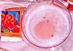
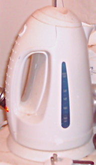
I took up my electric kettle, containing distilled water, and threw the switch. Seconds passed into minutes. Minutes passed into more minutes. Then, the water began boiling vigorously, and I applied one cup (8 fluid ounces) of this to the dish of powder, stirring it with a tablespoon. It seemed to take at least two minutes of continuous stirring, but the deceptively innocent-looking powder finally dissolved without the slightest scent of brimstone. As prescribed by the instructions on the box, I added a further 8 fluid ounces of cold water (from the tap of my kitchen sink), stirred briefly to mix, and placed the dish in the refrigerator to gel overnight.

I lay awake in bed for hours, wondering if I was doing the right thing. Was I insane? Did I not remember the tales of Jeckyll and Hyde? Of Doctor Frankenstein? Of Pons and Fleischman? What horrible fate was I setting myself up for? Finally, I dropped into a fitful slumber, disturbed only by dreams of amorphous orange demons stalking me to feast upon my soul…

Day broke, and this very afternoon I took the now solidified mass from the refrigerator. This was it. My last chance to avoid whatever hellish abuses this disturbingly orange substance had planned for me. But no…it was far too late to turn back now. I took up my spoon, and devoured every last bit of happy orange jiggliness.

This was approximately seven hours ago. In the intervening time, I have experienced the following symptoms: Occasional thirst, mild generalized anxiety about the near future, hunger, and an urge to write this blog post in a hyperbolic language more suited to an H.P. Lovecraft story than a scientific report. In other words…I appear to have been entirely unaffected, despite consuming an entire box of expired gelatin.

I’ve been taught that when hypothesis-testing, one considers the “null hypothesis”. That is, the hypothesis that would falsify the one that I’m starting with. In this case, it would be something to the effect of “I will suffer no harm whatsoever from eating expired JellO®”. Given the results in this experiment I must – in the tortured language of philosophical science – “fail to reject the null hypothesis”, because my results show no evidence whatsoever that I have suffered harm from eating expired gelatin. In other words, I cannot rationally cling to my original hypothesis as written, and must confess that perhaps expired instant gelatin still in intact packaging may, in fact, be harmless.

Ah, but I know what happens now. “Cad!”, you cry! “Fraud! Sham! This experiment is, like, totally bogus! This is not normal JellO® but a sugar-free impostor! And furthermore, this isn’t even JellO®-brand gelatin, but a cheap knock-off brand! How dare you, sir, feed us this crap, which proves nothing!”

I answer in two parts: Firstly, ladies and gentlemen who are my readers, I assure you that the contents of the less-famous brand and the official Kraft® Foods brand are essentially identical, and indeed, might conceivably have come from the same source. It’s common practice for one factory’s product to be shipped to multiple sellers who each offer it under their own label, as the wide variety of affected brands during the recent “salmonella peanut butter” scare demonstrated. And secondly: as it happens, I also have in my possession a box of JellO®-brand lime-flavored gelatin, WITH sugar, which although it lists no obvious “expiration date”, has a code stamped on the box indicating that it was originally packaged in late 2003, and therefore should have exceeded the expected 24-month shelf-life about the same time as today’s test subject did. I swear to you, dear readers, that I will repeat my experiment with this sample next.

Stay tuned: “Expired JellO II: Lime’s Revenge”, coming soon to a blog near you!

UPDATE: The Expired JellO® Saga continues here!

Okay, this is completely bogus…

The folks over at BBspot are callously spreading microbiological misinformation (click for full-size):

This is just plain irresponsible and utterly wrong. Surely everyone can see the obvious problem here, right?

When this happens, the flame is blue, not orange.

Uh, or so I’ve heard.

(If it isn’t obvious – what you do in this situation is calmly take the spreader out and set it on your nice, flameproof benchtop, and set something non-fragile and non-flammable on top of the flaming jar of alcohol, which will then go out quickly as the oxygen gets used up. All the labs I’ve been in lately use “canning” jars for the alcohol in this application, complete with lids which can be set on top to extinguish the flame.)

The Gram Stain Post to End All Gram Stain Posts

Gram stain, Gram stain, Gram stain! Bah. I think it’s time Microbiology grew up and moved out of Medicine’s basement.

Sure, the Gram stain[1] has its uses, but the procedure is grossly over-hyped. “[…]the most important stain in microbiology[…]”[2]! “[…]it is almost essential in identifying an unknown bacterium to know first whether it is Gram-positive or Gram-negative.”![3] “The Gram Stain reaction is an especially useful differentiating characteristic.[…]The Gram reaction turns out to be a property of fundamental importance for classifying bacteria phylogenetically as well as taxonomically.”![4] “[…]differentiates bacteria into two fundamental varieties of cells.”![5] “The Key to Microbiology“![6] [emphasis added…]

Bah! Sure, the Gram stain has its uses, but the hype it gets (even 125 years after its invention) is ridiculous. It’s worse than Harry Potter!

You really want to know what the Gram reaction tells you? Really? Okay, here it is:

A “Gram Positive” reaction tells you that your cells have relatively thick and intact cell walls

A “Gram Negative” reaction tells you that they don’t.

That’s it. That’s about all you can reliably infer from the Gram stain.

Previously, I put up a post describing what was my understanding of the conventional view of why the Gram stain works. Today, I’ll give you a much more detailed – and more correct – explanation of why it works as well as what its real significance is to identification of microbes. But first, a brief one-paragraph rant on why I think the Gram stain has such a hold on microbiology teaching.

I blame the fact that microbiology education is still largely in the shadow of medical technology education. When you artificially exclude the 99+% of organisms that aren’t associated with human diseases, the tiny number left do, indeed, seem to largely separate into two phylogenetic categories. Judging by what I’ve encountered thus far, it seems you get a lot of Proteobacteria (especially ?-Proteobacteria, like E.coli), which are “Gram-negative”. You also get a lot of Firmicutes (Bacillus, Streptococcus, Staphylococcus, etc.), and a couple of scattered Actinobacteria (Mycobacterium, for tuberculosis and leprosy, Corynebacterium for diptheria…). Both of these are considered “Gram-positive” (although if you use the standard procedure these days, the Mycobacteria may show no reaction at all). That’s, what, 3 phyla out of about 25 eubacterial and archael phyla? If we throw in Syphilis and Chlamydia, that’s still only 20% or so of the currently recognized prokaryotic phyla. If your microbiology classes assume everybody is training to be a medical technologist or clinical microbiologist, then the Gram stain becomes inflated in importance.

Enough of that – here’s a quick review of how the Gram stain works. Solutions of “Crystal Violet” (a purple dye) and Iodine are applied to cells fixed to a slide, where they soak in and precipitate in the cells. A “decolorizer” (usually ethanol) is applied to see if it will wash this dye precipitate out of the cells. A different, lighter-colored dye (such as safranin) is added so that the cells which DO have their dye washed out can be seen as well. In the end, “Gram positive” cells are a dark purple from the crystal violet/iodine that was not washed away, and “Gram negative” cells are not dark purple. (Usually they are pink, from the safranin, assuming that’s the dye used as the counterstain.)

Note that this does not differentiate cells into “two fundamental types” as is often claimed. You actually get four types: Groups of cells that are normally always “Gram positive”, Groups of cells that are normally always “Gram negative”, Groups of cells that are normally sometimes “Gram positive” and sometimes “Gram negative” (“Indeterminate”, or as I like to call it, “Gram-biguous”), and groups of cells that are normally NEITHER Gram-positive nor Gram-negative, like Mycoplasma, which aren’t dyed at all by the process. Incidentally, phylogenetically speaking, Mycoplasma is one of the “Gram positive” Firmicutes, just like Bacillus and Staphylococcus.

It’s kind of interesting to me that the Gram stain reaction has been such a mystery up until a century after its invention. What is it that makes “Gram positive” cells retain the dye while “Gram negative” ones don’t? Along the way, it seems like nearly every part of the bacterial cell was hypothesized to be the reason for the Gram reaction – lipids, carbohydrates, nucleic acids, “Magnesium ribonucleates”, and so forth. Davies et al, 1983, includes a table listing many of these and referencing historical papers making the claims. The fact that the reaction had something to do with the cell wall seems to go back quite a while, though the “Magnesium ribonucleates” idea doesn’t seem to have been entirely abandoned until the mid-1960’s[7]. It was also hypothesized that the “Gram positive” cells simply absorb more dye and therefore take longer to “decolorize”.

It turns out that “Gram-positive” cells actually don’t, necessarily, take up more dye than Gram negative ones. This was tested by taking a set concentration of bacterial cells and adding them to a set concentration of dye. After letting them soak, the samples were centrifuged to remove the bacteria, and the amount of dye found to be missing from the liquid was taken as the amount absorbed by the cells. They found that some Gram negative cells actually took up more dye than the Gram positives did. So much for that idea.[8]

Even relatively recently, I’ve seen it written that the bacterial cell wall, specifically, is what holds onto the stain, but even that turns out not to be true. Although the cell wall is the structure that seems to be responsible for the Gram reaction, in the late 1950’s it was demonstrated that it was not actually the staining of the cell wall that caused the reaction, but rather the ability of the cell wall to keep the decolorizer out of the cell.[9]

Apparently, the Crystal Violet/Iodine complex itself doesn’t even play a vital role. The complex apparently dissolves again more or less instantly as soon as the decolorizer touches it[10], and it’s even possible to differentiate “Gram positive” and “Gram negative” with simple stains like methylene blue or malachite green, if you’re clever about it[11]. The latter authors set up a clever test with crushed cell material, dye, and paper chromatography. They had the decolorizer soak into the paper, past a spot where dye-soaked cell material from Gram-positive and Gram-negative cells was placed, and watched for obvious differences in the amount of time it took the dye to be carried out by the decolorizer. Incidentally, my quick examination of this paper makes it look like cheaper 100% isopropyl alcohol (“rubbing alcohol”) might be slightly better than the standard 95% ethanol for Gram stains.

– INTERLUDE –

So, here we are at 1970 or so, and we already know that the Gram reaction is entirely based on how well the cell wall structure prevents organic solvents (like ethanol) from soaking into the cell to dissolve the dye complex. Yes, the mystery of why the Gram stain works in normal cells was largely solved by the Nixon era.
A few corners of the mystery remained, though. Why do “old” cultures of “Gram positive” cells often end up staining “Gram negative”, for example? Why do some kinds of cells seem to be sometimes Gram positive and sometimes Gram negative in the same culture? What, exactly, is really happening to the cell, deep down, during the staining process?

In 1983, the Gram Stain made the great technological leap into the 1930’s, when a variation of the technique was devised which allowed the Gram Stain to be observed by electron microscopy[12]. Using a funky platinum compound in place of iodine, the electron microscope reveals exactly where the dye complex is at any particular stage of the Gram stain process. Using this technique, it was possible to see how the decolorizer disrupts the outer membrane of classically-Gram-negative organisms and to see that the decolorizer potentially damages the cell wall and interior membrane, possibly allowing cell material to leak out (or decolorizer to get in and dissolve the dye complex). It was also seen that the dye complex permeates the entire cell, not just the cell wall.[13]

If you’ve been wondering about the sometimes-Gram-positive-sometimes-Gram-negative cells, the same technique was also used to investigate this. As suspected, it turns out that the “old cultures become Gram negative” problem is due to the cell walls breaking down as the culture ages. Bacteria are continuously, simultaneously, building up and tearing down their cell walls, in order to be able to grow and divide. As nutrients run out, the bacteria run out of material to rebuild cell walls, while the cell-wall degrading enzymes keep on chugging. Breaks in the cell wall occur, and through these breaks the decolorizer can get in and rapidly dissolve the dye. Actinobacteria can have a similar problem, but rather than only being in “old” cultures, apparently weaknesses appear briefly during cell division, and if a particular cell happens to be at this stage of growth when you stick it on a slide, heat-fix, and Gram stain it, the weakness at the septum where the division is occuring can crack and allow the decolorizer in, resulting in a “Gram negative” response even while surrounding cells of the same kind might still be “Gram positive”.[14]

This brings us to archaea and some eukaryotes (i.e. yeasts). Yeasts stain “Gram positive” normally. Although their cell walls are completely different chemically than bacterial cell walls, they are quite thick (microbially speaking). Poor, neglected Archaea seem to be all over the place in terms of Gram reaction. Since their Gram reaction doesn’t tend to correlate to any particular phylogenetic grouping[15], it seems nobody really pays much attention to their Gram stain reaction. On the other hand, and on the subject of “Gram-biguity”, I thought the investigation of Methanospirillum hungatei[16] was interesting. M.hungatei is an archaen that grows in chains. When Gram-stained, the cells on the ends of the chains are “Gram positive”, while the others have no Gram reaction at all. It turns out that the chains are covered by a sheath, and the only contact with the outside world is through thick “plugs” in the cells at the ends of the chains. These “plugs” act like thick cell walls, allowing the Gram stain dye material to soak in but excluding the decolorizer, while the sheath keeps the rest of the cells from soaking up any stain at all.

There you have it – a relatively detailed history and explanation for the Gram stain, and you didn’t even have to get through some obnoxious paywall to read it. Aren’t you lucky?

Comments, suggestions, and corrections, as always, are welcome.

[1] Gram, HC.”Ueber die isolirte Faerbung der Schizomyceten in Schnitt-und Trockenpraeparaten.” Fortschitte der Medicin. 1884 Vol. 2, pp 185-189.

[2] Popescu A, Doyle RJ. “The Gram stain after more than a century.” Biotech Histochem. 1996 May;71(3):145-51.

[3] Brock TD, Madigan MT, Martinko JM, Parker J. “Biology of Microorganisms (7th Edition).” 1994. Prentice Hall, Englewood Cliffs, NJ pg. 46

[4] ibid, pg. 715

[5] Beveridge TJ.”Use of the gram stain in microbiology.” Biotech Histochem. 2001 May;76(3):111-8.

[6] McClelland, Rosemary. “Gram’s stain: The key to microbiology – isolate identification method – Tutorial” Retrieved 20070810 from http://findarticles.com/p/articles/mi_m3230/is_4_33/ai_74268506/print

[7] Normore WM, Umbreit WW.”Ribonucleates and the Gram stain.” J Bacteriol. 1965 Nov;90(5):1500.

[8] BARTHOLOMEW JW, FINKELSTEIN H:”CRYSTAL VIOLET BINDING CAPACITY AND THE GRAM REACTION OF BACTERIAL CELLS.” J Bacteriol. 1954 Jun;67(6):689-91.

[9] BARTHOLOMEW JW, FINKELSTEIN H.”Relationship of cell wall staining to gram differentiation.” J Bacteriol. 1958 Jan;75(1):77-84.

[10] LAMANNA C, MALLETTE MF. “CHROMATOGRAPHIC ANALYSIS OF THE STATE OF ASSOCIATION OF THE DYE-IODINE COMPLEX IN DECOLORIZATION SOLVENTS OF THE GRAM STAIN.” J Bacteriol. 1964 Apr;87:965-6.

[11] Bartholomew JW, Cromwell T, Gan R.”Analysis of the Mechanism of Gram Differentiation by Use of a Filter-Paper
Chromatographic Technique.” J Bacteriol. 1965 Sep;90(3):766-77.

[12] Davies JA, Anderson GK, Beveridge TJ, Clark HC.”Chemical mechanism of the Gram stain and synthesis of a new electron-opaque marker for electron microscopy which replaces the iodine mordant of the stain.” J Bacteriol. 1983 Nov;156(2):837-45.

[13] Beveridge TJ, Davies JA.”Cellular responses of Bacillus subtilis and Escherichia coli to the Gram stain.” J Bacteriol. 1983 Nov;156(2):846-58.

[14] Beveridge TJ. “Mechanism of Gram Variability in Select Bacteria.” J Bacteriol. 1990 Mar;172(3):1609-20.

[15] Beveridge TJ, Schultze-Lam S. “The response of selected members of the archaea to the gram stain.” Microbiology. 1996 Oct;142 ( Pt 10):2887-95. (Abstract)

[16] Beveridge TJ, Sprott GD, Whippey P. “Ultrastructure, inferred porosity, and gram-staining character of Methanospirillum hungatei filament termini describe a unique cell permeability for this archaeobacterium.” J Bacteriol. 1991 Jan;173(1):130-40.
Simplify, Simplify…

DNA seems to have two main threats to its well-being once it’s extracted and purified.

  • Nucleases
  • Spontaneous Hydrolysis by water

Nucleases are the big one that everyone seems to mention. The seem to be fairly sturdy enzymes, and they’re everywhere (including fingertips – hence the need to wear gloves whenever you get near DNA samples…), and they “eat” DNA rapidly. Theoretically, you can destroy the enzymes with enough heat, but you still need to worry about them getting in every time you pop open your sample to get some out.

Apparently, DNA even in pure water can tend to slowly fall apart spontaneously. It doesn’t happen very fast, but bit by bit, it can undo the links between the individual nucleotides.

A common way to try to deal with nucleases is to add EDTA to the solution. Nucleases need magnesium ions dissolved in the water to do their job, and EDTA tightly binds to magnesium (and calcium). The idea is to “use up” any stray magnesium ions in the solution so that even if nucleases get in, they’re inactive because they have no magnesium available. That’s why you see EDTA in the recipes for so many DNA-related solutions. Of course – EDTA doesn’t permanently bind up all the magnesium – there’s always a tiny fraction that stays in the solution. So, although EDTA can drastically slow down any nucleases, it won’t actually stop them.

There are also some interesting chemicals which can be added to destroy all proteins (including nuclease enzymes). Guanidine Thiocyanate is one rather nasty chemical that does this. 2-mercaptoethanol is another. Various other detergents like CTAB may also denature any proteins. Since they don’t harm the DNA in the process, you could keep the DNA sample dissolved in a solution with these chemicals…but then you can’t do PCR with the sample as it is, since the protein-denaturing chemicals will also destroy any enzymes that you WANT, like DNA Polymerase, when you try to mix it into your reaction.

I think the latter option will be great for collecting field samples (in fact, it’s papers specifically on the subject of preserving samples in the field with CTAB and Guanidine Thiocyanate based solutions that I’m adapting from), but isn’t going to be real useful once I’ve got my DNA relatively purified. What to do, what to do…

Actually, I think the answer’s simpler than I originally expected. I’ll just dry the purified DNA out. No water – no hydrolysis…and no nuclease activity, either.

I could actually just leave it as a dried pellet in the bottom of a microcentrifuge tube, but that leaves the problem of taking only a little bit of it for processing rather than taking the whole thing, and I want to avoid reconstituting it and re-drying it repeatedly. I think a variation of the “dry the DNA on a piece of paper” process will be in order – then I can just cut off a small strip of the paper to get a portion of the DNA. It appears that you can actually dunk the DNA-impregnated bit of paper right into whatever solution you’re using (like a buffered polymerase-and-primers solution for PCR) and go for it.

Among the several references I found on this, here are two:
Kawai J, Hayashizaki Y: “DNA Book”; Genome Res. 2003 13: 1488-1495
Burgoyne LA: U.S. Patent #5496562 “Solid medium and method for DNA storage” (1996); U.S. Patent and Trademark Office, Washington D.C.

Colony PCR – because DNA extraction protocols suck.

If you’ve got a culture of a single type of bacteria and you want to identify it, the standard method is to figure out the sequence of one particular gene, the 16s rDNA gene. That is – it’s the gene which encodes the a piece of RNA that gets used by the ribosome in part of the process of “reading” which amino acids to link together to make a particular protein. This is something that every prokaryote known has, and parts of it are conserved, so they’re similar enough to compare, while other parts can vary a lot, providing enough “difference” to tell different organisms apart.

To figure out the sequence, you use PCR to “amplify” this particular gene, making lots of copies of it so that the sequencing machine can clearly see the signal from each part of the sequence. And before you can do that, you have to get the DNA out of the cell relatively intact.

That part can be a pain. There are lots of different ways people have come up with (and made special canned “kits” out of) – you can use chemicals to try to dissolve the cells and let all their guts (including the DNA) out, you can try to mash them up with tiny glass beads in a “bead-beating” machine, you can stick them in a blender, you can even just boil them for a while…then usually you go through several steps of centrifuging and mixing with different chemicals and then centrifuging again until you’ve hopefully finally got the DNA out and gotten rid of most of the other cell bits. And, hopefully, you haven’t accidentally chopped up the DNA too much to use in the process.

Fortunately, there’s a trick you can sometimes use, referred to as “Colony PCR”. In it, you literally just touch the top of your colony of cells and shake them off directly into the PCR tube. Then you just include an extra 5-10 minutes of 95°C heating to hopefully cook open enough of the cells to release DNA (and cook the cell’s enzymes to death so they don’t degrade the DNA and interfere with the PCR).

Not real reliable if you’re trying to do anything quantitative, like trying to figure out how many copies of a gene are in each cell, or trying to get an accurate estimate of how many cells of one type or another are in a mixed culture, but if you just need as much of a particular bit of DNA as you can get – such as for sequencing – a lot of people use this.

I just tried it on my Lambic isolates. Two of the 8 bacterial cultures worked beautifully. I’m pretty sure the problem with the other 6 was just the sheer amount of bacteria I ended up adding to the reaction – too much seems to “swamp” the PCR process and keep it from working. I’ll try it again this week. But it does seem to indicate that it works, at least.

E.coli – the “Microsoft” of the biotech world?

…by which I mean, it’s not always the best tool for the job, but everyone insists on always using it anyway, and has a variety of excuses for doing so…

Honestly – I’m trying to set up a clone library of 16s rDNA sequences using this kit. Never mind which kit it is – it actually does seem to work. I was just struck by the amount of hassle involved in shipping and storing the kit and it’s supply of “competent cells”.

When you get them, take them out of the dry-ice they’re shipped in and put them in the -80°C freezer immediately or they’ll die! Only thaw them carefully just before you use them, and do it on ice or they’ll die! Don’t heat-shock them for more than exactly 30 seconds or they’ll die! Once you’ve got them growing, you have to keep moving them to fresh selective media frequently or they’ll die! Or, you can carefully place them in the -80°C freezer…or they’ll die! Don’t look directly at them or they’ll die! (Do Not Taunt HappyFunCell!…)…

Seriously, running those gigantic -80° freezers can’t be cheap. Wouldn’t it be more convenient if you could grow up your transformant as an ordinary culture and just add your DNA samples and some kind of inducer chemical to make them take it up? Surely there must be some other organism that might be made to work like that.

Actually, it seems a number of the “Gram-positive” (firmicutes) organisms can enter a state of “natural competence”, where they naturally take up double-stranded DNA molecules from the environment. Bacillus subtilis is one. I’ve even seen references to “natural-competence” based protocols for transforming B.subtilis (or other Bacillus species, presumably) but it only seems to be in an out-of-print, $400 book.

Wouldn’t that be more convenient (using B.subtilis that is, not the $400 book)? Plus, when you wanted to store your transformed culture for later use, you could just heat the culture up to, what, about 55°C for 15 minutes or so (as I recall) then let it dry. The spores will contain whatever “bonus” plasmid DNA you added (if spores didn’t keep plasmids, then anthrax wouldn’t be such a danger…) and will last practically forever at room temperature. Mix the spores with some dried nutrient powder and seal them in a foil packet. Instant transformants, just add water!

But NOOOOO…..”But, everybody else uses E.coli, so I have to.” “They only make ‘BogoGen SuperMiniUltraKlone Kit 2000’ with E.coli, and we have to use that!” “But, nobody knows that other stuff, but everybody’s already familiar with E.coli!” “I’m a BogoGen Certified E.Coli Engineer, and I say everything else is just a toy and doesn’t work!” “All the books and stuff are about E.coli…”

Bah! Pathetic excuses. Anybody got a huge wad of venture capital to throw at me? The more I think about this, the more I think ‘untapped niche’…Heck, the electricity savings on not having to run a -80°C freezer constantly alone ought to qualify for a good “Fight Global Warming – Say ‘No!’ to E.coli!” marketing campaign…

Bonus perk: All the natto you can eat…

More Lambic pictures

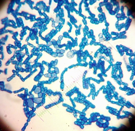
Ah, that’s better – a more traditional heat-fix/simple stain (using Methylene blue) shows my yeast isolates better:

Sally the maybe-Brettanomyces-type yeast
(“Sally”, a yeast that I suspect is a Brettanomyces-type yeast.)

Sam the...Saccharomyces-type yeast?
(“Sam” now looks awfully small…but more experienced observers than I am said that it could actually be a Saccharomyces-type yeast.)

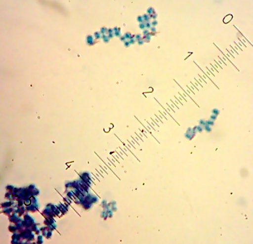
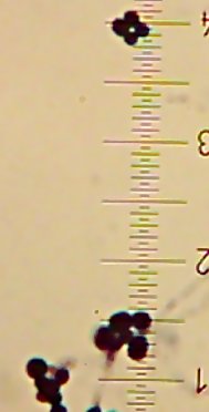
Lucy the possibly-PediococcusI also got two more Coccoid-Cluster-type Gram-positive bacterial isolates. The look pretty much the same under the microscope, though one had gooey wet, slightly larger colonies than the other’s smaller, hard-lump colonies. I see another one of those tetrads in the hard-lump-colony microscope image.

All told, I now have 10 isolates to check out. I’ve been given the go-ahead to try sequencing on the 8 bacterial isolates so hopefully I’ll be able to get a clear identity for Fred, Sid, Lisa, Lucy, BillyBob, JimBob, BettySue, and MarySue. Sally and Sam will have to wait for now, though I’m looking into ways to characterize them, too.