
“Ueber die isolirte Faerbung der Schizomyceten in Schnitt- und Trockenpraeparaten”

The Giant’s Shoulders blog carnival is coming up in two days, and I just realized I still haven’t gotten a post up for it yet. So, here it is.

I put up some quick reviews of several classic microbiology-methods papers for the previous edition of this blog carnival, but didn’t actually get around to putting up the one for what is almost certainly the most well-known microbiology technique: “The Gram Stain”. So, this post is about it:

Gram HC: “Ueber die isolirte Faerbung der Schizomyceten in Schnitt- und Trockenpraeparaten”; Fortschritte der Medicin; 1884; vol 2, pp 185-189

That’s “Regarding the Isolational(?) Coloring of Schizomycetes in Cut- [i.e. tissue sections] and Dried Preparations” in “Medical Progress”. The translation hosted by the American Society for Microbiology uses the word “Differential” where I’ve put “Isolational” – which is probably not quite right either but it’ll have to do for now – but I’ll get to that in a moment.

If you’ve ever been exposed to microbiology labwork before, you’ve almost certainly done or at least watched a procedure referred to as a “Gram stain”. In brief, you smear your sample with bacteria on a glass slide and bake it on, then you dump some purple stuff on it, them some brown stuff, then you rinse it briefly with alcohol, then you dump on some pink stuff, and then rinse it in water and look at it under a microscope. Bacteria that stay the original dark purple-blue color of the original purple/brown stuff are considered “Gram Positive”, and those that don’t instead appear the pink color of the last stain, and are considered “Gram Negative”. Many textbook authors and microbiology instructors will breathlessly proclaim that the Gram Stain reveals two “fundamental” categories of bacteria, but I’ll spare you my rant about that.

Properly speaking, this isn’t actually Gram’s stain, as described in his original paper. The modern variations that we’re all taught in microbiology class were developed later, and I believe they are nowadays based mainly on Victor Burke’s 1922 paper on the subject[1].

Regarding the title of the paper: “schizomycete” is what they used to call most kinds of bacteria. “Mycete” meaning “fungus”, as bacteria were assumed to be “plants without chlorophyll” just like molds and mushrooms, and “Schizo-” meaning “split in two”, since bacteria reproduce by splitting into two cells rather than by producing spores like “other” fungi. I say “most” because things like cyanobacteria (“blue-green algae”) or Green Sulfur Bacteria would have been referred to as “Schizophyta” (“fission-plants”). What Gram was originally trying to do wasn’t to differentiate one kind of bacteria from another, either, but to make it easy to tell bacteria from from the nuclei of cells in bacteria-infected tissue.

For that matter, Gram was really metaphorically standing on the shoulders of Koch and Erhlich, as he was building on their technique for staining “tubercle bacteria” – that is, tuberculosis-causing members of the genus Mycobacterium. Gram mentions that you need to stain this type of bacteria for the “usual” 12-24 hours to make this work, incidentally, as opposed to a few minutes for other “schizomycetes”. This suggests that you are expected to have some idea of what you’re going to find before you use the stain, as opposed to the modern implementation which is supposed to tell you something about what kind of bacteria you’re finding.

Still, Gram does report that some bacteria take the stain and some don’t, giving us a preview of the “differential” character of the modern version. He specifically notes typhoid and some causes of bronchial pneumonia fail to hold the stain. Given that Typhoid Fever is caused by a strain of the “Gram-negative” butt-bacter Salmonella enterica, and there are a number of “Gram negative” bacteria as well as “Gram positive” that can cause pneumonia, this makes sense. He also does mention the use of Bismarck Brown R a.k.a. Vesuvine as a counterstain in order to make the nuclei of the infected cells brown in contrast to the dark blue of the infectious bacteria in the tissue.

For much of the century-and-a-quarter since Gram’s publication, the question of why the Gram stain works was thoroughly investigated, and even today I occasionally hear or read assertions to the effect that the Gram Stain isn’t well understood. I disagree with this just as I think its importance to bacterial identification is grossly overblown, and if you want to know why, I have a previous post all about why the Gram stain works and how we know. You may or may not also be interested in an older post regarding whether or not “acid-fast” bacteria like the ones that cause tuberculosis (which don’t stain at all when using the modern version of the Gram stain) are “Gram Positive” or not. As always, if you spot any errors or have any questions, please let me know…

[1] Burke V: “Notes on the Gram Stain with Description of a New Method.” J Bacteriol. 1922 Mar;7(2):159-82.
SHENANIGANS! Caffeine is our FRIEND!

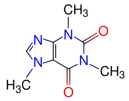
Our new Asylum has real internet finally now and we’re getting settled in. The Houston area here is one of the most hot and humid areas of the US. All hot and sweaty. So of course I’ve been advised that my favorite psychotropic substance – 1,3,7-trimethylxanthine [“caffeine” for party-poopers who aren’t into the fancier names] – is no longer my friend, because it’s a diuretic that’ll dehydrate me, right?

NO! Shenanigans! Caffeine is our FRIEND! And that stuff about it being a diuretic? CRAP! LIES AND SLANDER!

But don’t just take my word for it. After all, humans are a bunch of freakish multicellular soft-celled eukaryotes, and I normally focus on normal organisms like bacteria, archaea, and yeasts. So, let’s ask some real human-physiology type scientists and check out their official peer-reviewed findings:

Armstrong LE, Pumerantz AC, Roti MW, Judelson DA, Watson G, Dias JC, Sokmen B, Casa DJ, Maresh CM, Lieberman H, Kellogg M: “Fluid, electrolyte, and renal indices of hydration during 11 days of controlled caffeine consumption.”; Int J Sport Nutr Exerc Metab. 2005 Jun;15(3):252-65.

“[…]The following variables were unaffected (P > 0.05) by different caffeine doses on days 1, 3, 6, 9, and 11 and were within normal clinical ranges: body mass, urine osmolality, urine specific gravity, urine color, 24-h urine volume, 24-h Na+ and K+ excretion, 24-h creatinine, blood urea nitrogen, serum Na+ and K+, serum osmolality, hematocrit, and total plasma protein. Therefore, C0, C3, and C6 exhibited no evidence of hypohydration.[…]”

Abstract on Pubmed

Armstrong LE, Casa DJ, Maresh CM, Ganio MS: “Caffeine, fluid-electrolyte balance, temperature regulation, and exercise-heat tolerance.” Exerc Sport Sci Rev. 2007 Jul;35(3):135-40.

“[…]This review, contrary to popular beliefs, proposes that caffeine consumption does not result in the following: (a) water-electrolyte imbalances or hyperthermia and (b) reduced exercise-heat tolerance.”

(Review article, apparently – Abstract on Pubmed)

Del Coso J, Estevez E, Mora-Rodriguez R: “Caffeine effects on short-term performance during prolonged exercise in the heat.” Med Sci Sports Exerc. 2008 Apr;40(4):744-51.

“[…]RESULTS: Without fluid replacement (NF and NF + CAFF), subjects were dehydrated by 3.8 +/- 0.3%[…]CONCLUSION: During prolonged exercise in the heat, caffeine ingestion (6 mg.kg body weight) maintains MVC and increases PMAX despite dehydration and hyperthermia. When combined with water and carbohydrate, caffeine ingestion increases maximal leg force by increasing VA (i.e., reducing central fatigue).”

(“NF” = “No Fluid replacement” – the “dehydration” mentioned here is due to exercising in the heat, and doesn’t appear to be related to whether the test subjects consumed caffeine or not)

Abstract on Pubmed

Scott D, Rycroft JA, Aspen J, Chapman C, Brown B:”The effect of drinking tea at high altitude on hydration status and mood.” Eur J Appl Physiol. 2004 Apr;91(4):493-8. Epub 2004 Feb 11.

“[…]Several markers of hydration status were also taken immediately pre and post each condition, including measures of urine specific gravity, urine electrolyte balance (K+, Na+), and urine colour. None of these measures indicated a difference in hydration status as a result of the dietary intervention in either the control or tea condition.[…]”

(In this study, the tea was the only caffeine-containing substance involved. The study group’s caffeine came solely from the tea. The control group got no caffeine at all.)

Abstract on pubmed

Paluska SA: “Caffeine and exercise.” Curr Sports Med Rep. 2003 Aug;2(4):213-9.

“[…]It[caffeine] is relatively safe and has no known negative performance effects, nor does it cause significant dehydration or electrolyte imbalance during exercise.[…]”

Abstract on Pubmed

Grandjean AC, Reimers KJ, Bannick KE, Haven MC.: “The effect of caffeinated, non-caffeinated, caloric and non-caloric beverages on hydration.” J Am Coll Nutr. 2000 Oct;19(5):591-600.

“[…]This preliminary study found no significant differences in the effect of various combinations of beverages on hydration status of healthy adult males.[…]”

Pubmed entry – full text available

See? Oh, I know what you’re going to say next – “But, like, dude! When I drink my Venti Mocha Crappucino [note: Link goes to “Foamy the Squirrel”, who is a bit of a pottymouth, ranting about the “Tall/Grande/Venti” nonsense.  It amused me.] or a can of Jolt Ultra I have to take a major whiz a little while later! Isn’t that ‘cuz of the caffeine?” Well, no, it isn’t. It’s because you just drank a bunch of liquid. Duh.

So, you see, caffeine really is our friend. Be nice to caffeine. But don’t feed it to your yeast in the presence of benzoic acid because it’ll kill them. See? I managed to turn this into a segue back to the stuff I was talking about before the whole “buy a house in Texas” thing started interfering. Stay tuned…

What really counts as a “microbe”?

Just a brief pre-post before the main one I’ve got brewing now (which will be posted either later today or tomorrow).

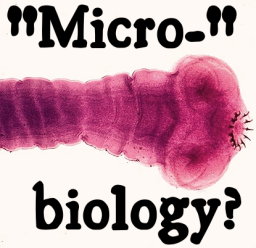
A tapeworm: Since when does 30-36 feet long count as 'micro'???Microbiology is the dominating topic of this particular blog, but I don’t think I’ve ever addressed what I consider to really count as “micro”biology. This isn’t necessarily an obvious topic. My old “Microbiology” book from 8 years ago, plus the textbook from last year’s “Pathogenic Microbiology” class both contained large sections discussing organisms that are visible without a microscope. Heck, the “Pathogenic Microbiology” text even had a whole section on spider and insect bites. And, tapeworms? Since when is “over 30 feet long” considered “micro”? As I like to say: It’s time for Microbiology to grow up and move out of Medicine’s basement.

So: Here are the defining features of what I consider to be a “microbe”, at least for purposes of what I tend to discuss here on the blog:

  • Obvious: the organism cannot be effectively examined visually without a microscope and individual organisms can virtually never be observed by the “naked eye”.
  • In nature, a full normal population of a microbe can and will develop from a single live cell, and isolated individual cells are reasonably commonly observed.
  • Microbes do not “eat”.

It’s that last point that prompted me to write this post, mainly because it’s such an important part of why microbes work and how they affect their surroundings, especially when it comes to food microbes. What I mean by “do not eat” is that they are incapable of taking large (microbially speaking) chunks of material into themselves to use. Any cell nutrient for a microbe must be in the form of small molecules, like sugars, small peptides or individual amino acids, and so on that can be easily transported across the cell membranes and through the cell wall where applicable.

The importance of this is that for a microbe to grow on a complicated substance like meat or bread (for example), they have to excrete specialized enzymes that break down the substances out in the environment into simpler components like sugars or small peptides. If a microbe cannot secrete a protein-digesting “protease” enzyme, it can be surrounded by tasty, nutritious proteins and still starve to death. If a microbe can’t secrete an amylase (starch-digesting) enzyme, it doesn’t matter that starch is made of nice yummy glucose molecules because they’re all wadded up into long chains of starch that the microbe can’t get at.

And that, finally, is important because it brings up issues of growing multiple microbes together to accomplish something. Sake, for example, is made by fermenting rice, but rice is made primarily of starch. Saccharomyces yeasts don’t make amylases, so in order to make sake, you also have to add a kind of mold (Aspergillus oryzae, one of the types of white-mold-with-little-black-specks that you may see growing on the bread you’ve left sitting around for too long). A. oryzae is also a microbe and therefore can’t “eat”, but it does produce amylase. Since the amylase is breaking down the starches outside of the cells, this means the released glucose is also available for the yeast to use.

Admittedly, my definition above isn’t perfect. On the one hand, it leaves out protozoa (like amoebae and the well-known Paramecium, both of which actually do take in “chunks” of food, but both of which most people would normally consider to be “microbes”. It also leaves IN things like mushrooms, which are not usually thought of as being “microbes” by people who aren’t microbiologists. And, of course, it leaves me with no excuse not to go and learn something about eukaryotic (“plant”) algae (as opposed to bacteria-algae, a.k.a. cyanobacteria) and diatoms. Suggestions for updating my definition may be left in the comments…

Just something that came up while I was assembling what will be the next post. Stay tuned.